Human CD1a APC-conjugated Antibody
R&D Systems, part of Bio-Techne | Catalog # FAB7076A


Conjugate
Catalog #
Key Product Details
Species Reactivity
Human
Applications
Flow Cytometry
Label
Allophycocyanin (Excitation = 620-650 nm, Emission = 660-670 nm)
Antibody Source
Monoclonal Mouse IgG1 Clone # 703217
Product Specifications
Immunogen
Mouse myeloma cell line NS0-derived recombinant human CD1a
Asp19-Val300 (predicted)
Accession # P06126
Asp19-Val300 (predicted)
Accession # P06126
Specificity
Detects human CD1a in direct ELISAs.
In direct ELISAs, less than 5%
cross-reactivity with recombinant human (rh) CD1d is observed and no
cross-reactivity with with rhCD1b or rhCD1e is observed.
Clonality
Monoclonal
Host
Mouse
Isotype
IgG1
Scientific Data Images for Human CD1a APC-conjugated Antibody
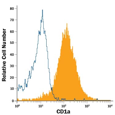
Detection of CD1a in MOLT‑4 Human Cell Line by Flow Cytometry.
MOLT-4 human acute lymphoblastic leukemia cell line was stained with Mouse Anti-Human CD1a APC-conjugated Monoclonal Antibody (Catalog # FAB7076A, filled histogram) or isotype control antibody (Catalog # IC002A, open histogram). View our protocol for Staining Membrane-associated Proteins.Applications for Human CD1a APC-conjugated Antibody
Application
Recommended Usage
Flow Cytometry
10 µL/106 cells
Sample: MOLT‑4 human acute lymphoblastic leukemia cell line
Sample: MOLT‑4 human acute lymphoblastic leukemia cell line
Formulation, Preparation, and Storage
Purification
Protein A or G purified from hybridoma culture supernatant
Formulation
Supplied in a saline solution containing BSA and Sodium Azide.
Shipping
The product is shipped with polar packs. Upon receipt, store it immediately at the temperature recommended below.
Stability & Storage
Store the unopened product at 2 - 8° C. Do not use past expiration date. Protect from light.
Background: CD1a
Long Name
T Cell Surface Glycoprotein CD1a
Alternate Names
CD1a, FCB6, HTA1, R4, T6
Entrez Gene IDs
909 (Human)
Gene Symbol
CD1A
UniProt
Additional CD1a Products
Product Documents for Human CD1a APC-conjugated Antibody
Product Specific Notices for Human CD1a APC-conjugated Antibody
For research use only
Loading...
Loading...
Loading...
Loading...
Loading...