Human CD40/TNFRSF5 Antibody
R&D Systems, part of Bio-Techne | Catalog # MAB6321


Key Product Details
Species Reactivity
Validated:
Cited:
Applications
Validated:
Cited:
Label
Antibody Source
Product Specifications
Immunogen
Glu21-Arg193
Accession # P25942
Specificity
Clonality
Host
Isotype
Endotoxin Level
Scientific Data Images for Human CD40/TNFRSF5 Antibody
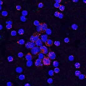
CD40/TNFRSF5 in Human PBMCs.
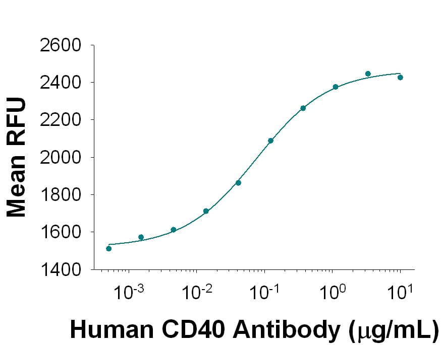
CD40/TNFRSF5 was detected in immersion fixed human peripheral blood mononuclear cells (PBMCs) using Mouse Anti-Human CD40/TNFRSF5 Monoclonal Antibody (Catalog # MAB6321) at 10 µg/mL for 3 hours at room temperature. Cells were stained using the NorthernLights™ 557-conjugated Anti-Mouse IgG Secondary Antibody (red; Catalog # NL007) and counterstained with DAPI (blue). Specific staining was localized to cell surfaces and cytoplasm. View our protocol for Fluorescent ICC Staining of Non-adherent Cells.Human CD40/TNFRSF5 Antibody Stimulates Cell Proliferation in Human B Cells.
Mouse Anti-Human CD40/TNFRSF5 Monoclonal Antibody (Catalog # MAB6321) stimulates human B cell proliferation in the presence of Recombinant Human IL-4 (Catalog # 204-IL) in a dose-dependent manner, as measured by Resazurin (Catalog # AR002). The ED50 for this effect is typically 0.035-0.175 µg/mLDetection of CD40/TNFRSF5 in PBMC lymphocytes by Flow Cytometry
PBMC lymphocytes were stained with Mouse Anti-Human CD19 APC-conjugated Monoclonal Antibody (Catalog # FAB4867A) and either (A) Mouse Anti-Human CD40/TNFRSF5 Monoclonal Antibody (Catalog # MAB6321) or (B) isotype control antibody (Catalog # MAB0041) followed by Phycoerythrin-conjugated Anti-Mouse IgG Secondary Antibody (Catalog # F0102B). View our protocol for Staining Membrane-associated Proteins.Applications for Human CD40/TNFRSF5 Antibody
Agonist Activity
CyTOF-ready
Flow Cytometry
Sample: Human whole blood CD19+ B cells
Immunocytochemistry
Sample: Immersion fixed human peripheral blood mononuclear cells (PBMCs)
Reviewed Applications
Read 1 review rated 4 using MAB6321 in the following applications:
Formulation, Preparation, and Storage
Purification
Reconstitution
Formulation
Shipping
Stability & Storage
- 12 months from date of receipt, -20 to -70 °C as supplied.
- 1 month, 2 to 8 °C under sterile conditions after reconstitution.
- 6 months, -20 to -70 °C under sterile conditions after reconstitution.
Background: CD40/TNFRSF5
CD40 is a type I transmembrane glycoprotein belonging to the TNF receptor superfamily. The mature hCD40 consists of a 172 amino acid (aa) extracellular domain, a 22 aa transmembrane region and a 62 aa cytoplasmic domain (1). Human and mouse CD40 share 62% aa identity. CD40 is expressed in B cells, follicular dendritic cells, dendritic cells, activated monocytes, macrophages, endothelial cells, vascular smooth muscle cells, and several tumor cell lines (2). The extracellular domain has the cysteine-rich repeat regions, which are characteristic for many of the receptors of the TNF superfamily. Interaction of CD40 with its ligand, CD40L, leads to aggregation of CD40 molecules, which in turn interact with cytoplasmic components to initiate signaling pathways. Early studies on the CD40-CD40L system revealed its role in humoral immunity. Interaction between CD40L on T cells and CD40 on B cells stimulated B cell proliferation and provided the signal for immunoglobulin isotype switching (3). Mutations in the CD40L gene, which resulted in a CD40L molecule unable to interact with CD40, are responsible for the hyper-IgM syndrome (4). Cross-linking of CD40 with antibodies or by CD40 binding to CD40L produces cell type-specific responses which include costimulation and induction of proliferation, induction of cytokine production, rescue from apoptosis, and upregulation of adhesion molecules (5). Some of the early events of intracellular signaling by the
CD40‑CD40L system include the association of the CD40 with TRAFs and the activation of various kinases (6‑8).
References
- Torres, R.M. and E.A. Clark (1992) J. Immunol. 148:620.
- Schonbeck, U. et al. (1997) J. Biol. Chem. 272:19569.
- Armitage, R.J. et al. (1993) J. Immunol. 150:3671.
- Callard, R.E. et al. (1993) Immunol. Today 14:559.
- Stout, R.D. and J. Suttles (1996) Immunol. Today 17:487.
- Pullen, S.S. et al. (1999) Biochemistry 38:10168.
- Faris, M. et al. (1994) J. Exp. Med. 179:1923.
- Hanissian, S.H. and R.S Geha (1997) Immunity 6:379.
Alternate Names
Gene Symbol
UniProt
Additional CD40/TNFRSF5 Products
Product Documents for Human CD40/TNFRSF5 Antibody
Product Specific Notices for Human CD40/TNFRSF5 Antibody
For research use only

