Human CD72 Antibody
R&D Systems, part of Bio-Techne | Catalog # MAB5405


Conjugate
Catalog #
Key Product Details
Species Reactivity
Human
Applications
CyTOF-ready, Flow Cytometry, Immunocytochemistry
Label
Unconjugated
Antibody Source
Monoclonal Mouse IgG2A Clone # 982405
Product Specifications
Immunogen
Chinese hamster ovary cell line CHO-derived recombinant human CD72
Arg117-Asp359
Accession # P21854
Arg117-Asp359
Accession # P21854
Specificity
Detects human CD72 in direct ELISAs.
Clonality
Monoclonal
Host
Mouse
Isotype
IgG2A
Scientific Data Images for Human CD72 Antibody
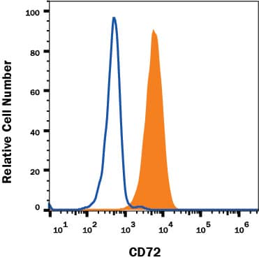
Detection of CD72 in Human Ramos Cell Line by Flow Cytometry.
Human Ramos Burkitt's lymphoma cell line was stained with Mouse Anti-Human CD72 Monoclonal Antibody (Catalog # MAB5405, filled histogram) or Mouse IgG2A Isotype Control (Catalog # MAB003, open histogram) followed by APC-conjugated Anti-Mouse IgG Secondary Antibody (Catalog # F0101B). View our protocol for Staining Membrane-associated Proteins.CD72 in Ramos Human Cell Line.
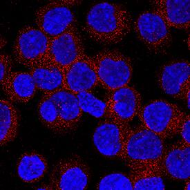
CD72 was detected in immersion fixed Ramos human Burkitt's lymphoma cell line using Mouse Anti-Human CD72 Monoclonal Antibody (Catalog # MAB5405) at 8 µg/mL for 3 hours at room temperature. Cells were stained using the NorthernLights™ 557-conjugated Anti-Mouse IgG Secondary Antibody (red; Catalog # NL007) and counterstained with DAPI (blue). Specific staining was localized to cytoplasm. View our protocol for Fluorescent ICC Staining of Non-adherent Cells.Applications for Human CD72 Antibody
Application
Recommended Usage
CyTOF-ready
Ready to be labeled using established conjugation methods. No BSA or other carrier proteins that could interfere with conjugation.
Flow Cytometry
0.25 µg/106 cells
Sample: Ramos Human Burkitt's Lymphoma Cell Line
Sample: Ramos Human Burkitt's Lymphoma Cell Line
Immunocytochemistry
8-25 µg/mL
Sample: Immersion fixed Ramos human Burkitt's lymphoma cell line
Sample: Immersion fixed Ramos human Burkitt's lymphoma cell line
Formulation, Preparation, and Storage
Purification
Protein A or G purified from hybridoma culture supernatant
Reconstitution
Reconstitute at 0.5 mg/mL in sterile PBS. For liquid material, refer to CoA for concentration.
Formulation
Lyophilized from a 0.2 μm filtered solution in PBS with Trehalose. *Small pack size (SP) is supplied either lyophilized or as a 0.2 µm filtered solution in PBS.
Shipping
Lyophilized product is shipped at ambient temperature. Liquid small pack size (-SP) is shipped with polar packs. Upon receipt, store immediately at the temperature recommended below.
Stability & Storage
Use a manual defrost freezer and avoid repeated freeze-thaw cycles.
- 12 months from date of receipt, -20 to -70 °C as supplied.
- 1 month, 2 to 8 °C under sterile conditions after reconstitution.
- 6 months, -20 to -70 °C under sterile conditions after reconstitution.
Background: CD72
References
- Nykimbeng-Takwi, E. and S.P. Chapoval (2011) Immunol. Res. 50:10.
- Von Hoegen, I. et al. (1990) J. Immunol. 144:4870.
- Nakayama, E. et al. (1989) Proc. Natl. Acad. Sci. 86:1352.
- Alcon, V.L. et al. (2009) Eur. J. Immunol. 39:826.
- Ishida, I. et al. (2003) Int. Immunol. 15:1027.
- Kataoka, T.R. et al. (2010) J. Immunol. 184:2468.
- Van de Velde, H. et al. (1991) Nature 351:662.
- Luo, W. et al. (1992) J. Immunol. 148:1630.
- Kumanogoh, A. et al. (2000) Immunity 13:621.
- Kumanogoh, A. et al. (2005) Int. Immunol. 17:1277.
- Li, D. H.-H. et al. (2008) Arthritis Rheum. 58:3192.
- Wu, H.-J. et al. (2001) J. Immunol. 167:1263.
- Mizrahi, S. et al. (2007) PLoS ONE 9:e818.
Alternate Names
CD72, Ly-19.2, Ly-32.2, Lyb-2
Gene Symbol
CD72
UniProt
Additional CD72 Products
Product Documents for Human CD72 Antibody
Product Specific Notices for Human CD72 Antibody
For research use only
Loading...
Loading...
Loading...
Loading...
Loading...
