Human Cyclin B2 Antibody
R&D Systems, part of Bio-Techne | Catalog # AF6204

Key Product Details
Species Reactivity
Applications
Label
Antibody Source
Product Specifications
Immunogen
Met1-Lys101
Accession # O95067
Specificity
Clonality
Host
Isotype
Scientific Data Images for Human Cyclin B2 Antibody
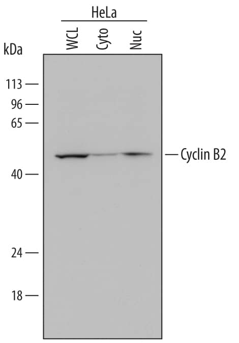
Detection of Human Cyclin B2 by Western Blot.
Western blot shows lysates of HeLa human cervical epithelial carcinoma cell line. Gels were loaded with 30 µg of whole cell lysate (WCL), 20 µg of cytoplasmic (Cyto), and 10 µg of nuclear extracts (Nuc). PVDF Membrane was probed with 1 µg/mL of Sheep Anti-Human Cyclin B2 Antigen Affinity-purified Polyclonal Antibody (Catalog # AF6204) followed by HRP-conjugated Anti-Sheep IgG Secondary Antibody (Catalog # HAF016). A specific band was detected for Cyclin B2 at approximately 48-54 kDa (as indicated). This experiment was conducted under reducing conditions and using Immunoblot Buffer Group 1.Cyclin B2 in Human Colon.
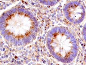
Cyclin B2 was detected in immersion fixed paraffin-embedded sections of human colon using Sheep Anti-Human Cyclin B2 Antigen Affinity-purified Polyclonal Antibody (Catalog # AF6204) at 15 µg/mL overnight at 4 °C. Before incubation with the primary antibody, tissue was subjected to heat-induced epitope retrieval using Antigen Retrieval Reagent-Basic (Catalog # CTS013). Tissue was stained using the Anti-Sheep HRP-DAB Cell & Tissue Staining Kit (brown; Catalog # CTS019) and counterstained with hematoxylin (blue). Specific staining was localized to epithelial cells in mucosal glands. View our protocol for Chromogenic IHC Staining of Paraffin-embedded Tissue Sections.Applications for Human Cyclin B2 Antibody
Immunohistochemistry
Sample: Immersion fixed paraffin-embedded sections of human colon
Western Blot
Sample: HeLa human cervical epithelial carcinoma cell line
Formulation, Preparation, and Storage
Purification
Reconstitution
Formulation
Shipping
Stability & Storage
- 12 months from date of receipt, -20 to -70 °C as supplied.
- 1 month, 2 to 8 °C under sterile conditions after reconstitution.
- 6 months, -20 to -70 °C under sterile conditions after reconstitution.
Background: Cyclin B2
Cyclin B2 (also CCNB2 and G2/mitotic-specific cyclin-B2) is a 48-54 kDa member of the Cyclin AB subfamily, cyclin family of proteins. It is widely expressed and associates with CDK1 providing substrate specificity to a phosphorylating complex. A phosphorCDK1:Cyclin B2 complex is associated with the Golgi apparatus, where it contributes to Golgi fragmentation during mitosis. It is also possible that Cyclin B2 can substitute for Cyclin B1 during the early stages of mitosis. Human Cyclin B2 is 398 amino acids (aa) in length. It contains two cyclin box folds (aa 201-290 and 298-383) and two substrate binding sites (aa 165-254 and 264-347). Phosphorylation occurs at Ser92, Thr94, Ser99, Ser204, Ser392 and Ser398. Over aa 1-101, human Cyclin B2 shares 79% aa identity with mouse Cyclin B2.
Alternate Names
Gene Symbol
UniProt
Additional Cyclin B2 Products
Product Documents for Human Cyclin B2 Antibody
Product Specific Notices for Human Cyclin B2 Antibody
For research use only

