Human Cytokeratin 19 Antibody
R&D Systems, part of Bio-Techne | Catalog # MAB35063


Key Product Details
Species Reactivity
Applications
Label
Antibody Source
Product Specifications
Immunogen
Gln311-Lys370
Accession # P08727
Specificity
Clonality
Host
Isotype
Scientific Data Images for Human Cytokeratin 19 Antibody
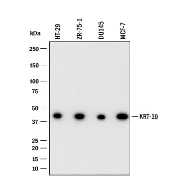
Detection of Cytokeratin 19 by Western Blot.
Western blot shows lysates of HT-29 human colon adenocarcinoma cell line, ZR-75-1 human breast cancer cell line, DU145 human prostate carcinoma cell line, and MCF-7 human breast cancer cell line. PVDF membrane was probed with 1 µg/mL of Mouse Anti-Human Cytokeratin 19 Monoclonal Antibody (Catalog # MAB35063) followed by HRP-conjugated Anti-Mouse IgG Secondary Antibody (HAF018). A specific band was detected for Cytokeratin 19 at approximately 40 kDa (as indicated). This experiment was conducted under reducing conditions and using Western Blot Buffer Group 1.Cytokeratin 19 in RT-4 Human Cell Line.
Cytokeratin 19 was detected in immersion fixed RT-4 human urinary bladder transitional cell papilloma cell line (positive staining) and THP-1 human acute monocytic leukemia cell line (negative staining) using Mouse Anti-Human Cytokeratin 19 Monoclonal Antibody (Catalog # MAB35063) at 8 µg/mL for 3 hours at room temperature. Cells were stained using the NorthernLights™ 557-conjugated Anti-Mouse IgG Secondary Antibody (red; NL007) and counterstained with DAPI (blue). Specific staining was localized to cytoplasm. Staining was performed using our protocol for Fluorescent ICC Staining of Non-adherent Cells.Cytokeratin 19 in MCF-7 Human Cell Line.
Cytokeratin 19 was detected in immersion fixed MCF-7 human breast cancer cell line (positive staining) and THP-1 human acute monocytic leukemia cell line (negative staining) using Mouse Anti-Human Cytokeratin 19 Monoclonal Antibody (Catalog # MAB35063) at 8 µg/mL for 3 hours at room temperature. Cells were stained using the NorthernLights™ 557-conjugated Anti-Mouse IgG Secondary Antibody (red; NL007) and counterstained with DAPI (blue). Specific staining was localized to cytoplasm. Staining was performed using our protocol for Fluorescent ICC Staining of Non-adherent Cells.Applications for Human Cytokeratin 19 Antibody
Immunocytochemistry
Sample: Immersion fixed RT‑4 human urinary bladder transitional cell papilloma cell line and MCF‑7 human breast cancer cell line
Immunohistochemistry
Sample: Immersion fixed paraffin-embedded sections of human prostate
Intracellular Staining by Flow Cytometry
Sample: MCF-7 breast carcinoma cell line fixed with Flow Cytometry Fixation Buffer (Catalog # FC004) and permeabilized with Flow Cytometry Permeabilization/Wash Buffer I (Catalog # FC005)
Western Blot
Sample: HT‑29 human colon adenocarcinoma cell line, ZR‑75-1 human breast cancer cell line, DU145 human prostate carcinoma cell line, and MCF‑7 human breast cancer cell line
Formulation, Preparation, and Storage
Purification
Reconstitution
Formulation
Shipping
Stability & Storage
- 12 months from date of receipt, -20 to -70 °C as supplied.
- 1 month, 2 to 8 °C under sterile conditions after reconstitution.
- 6 months, -20 to -70 °C under sterile conditions after reconstitution.
Background: Cytokeratin 19
Cytokeratin 19 (Keratin, type I cytoskeletal 19; also KRT-19, CK19 and Keratin-19) is a 40-45 kDa, acidic Class I keratin member of the intermediate filament family of proteins. Individual keratins are always expressed in tandem with a second keratin, and these are found in all epithelial cells. The class I KRT-19 heterodimerizes/polymerizes with 50-52 kDa class II KRT-8 (plus KRT-5 and -7) to form 8-10 nm filaments in epidermal stem cells, secretory gland (sweat; mammary; bile duct) simple epithelium, and neuroendocrine epidermal Merkel cells. It may represent a viable marker for skin stem cells. In skin, Cytokeratin 19 forms filaments in the fetal epithelium, and then progressively decreases with age, being virtually absent by age 17. Human Cytokeratin 19 is 400 amino acids (aa) in length. It contains an N-terminal "head" region (aa 1-79) and a subsequent "rod" region (aa 80-387), but is absent a typical C-terminal tail region. Cytokeratin 19 possesses at least 5 utilized phosphorylation sites plus one acetylated Lys residue. Based on other keratins, and the presence of an Asp at position 238, there may be caspase cleavage-generated isoforms. Full length human Cytokeratin 19 (aa 2-400) shares 82% aa sequence identity with mouse Cytokeratin 19.
Alternate Names
Gene Symbol
UniProt
Additional Cytokeratin 19 Products
Product Documents for Human Cytokeratin 19 Antibody
Product Specific Notices for Human Cytokeratin 19 Antibody
For research use only


