Human DC-SIGN/CD209 Fluorescein-conjugated Antibody
R&D Systems, part of Bio-Techne | Catalog # FAB161F


Key Product Details
Species Reactivity
Validated:
Cited:
Applications
Validated:
Cited:
Label
Antibody Source
Product Specifications
Immunogen
Specificity
Clonality
Host
Isotype
Scientific Data Images for Human DC-SIGN/CD209 Fluorescein-conjugated Antibody
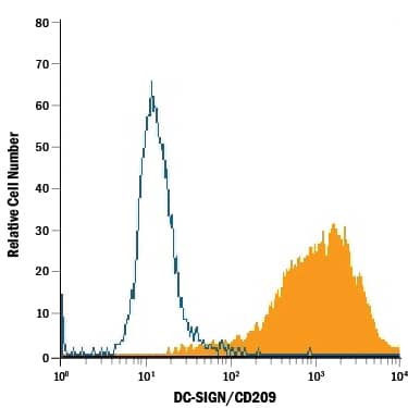
Detection of DC-SIGN/CD209 in NIH-3T3 Mouse Cell Line Transfected with Human DC-SIGN/CD209 by Flow Cytometry.
NIH-3T3 mouse embryonic fibroblast cell line transfected with human DC-SIGN/CD209 was stained with Mouse Anti-Human DC-SIGN/CD209 Fluorescein-conjugated Monoclonal Antibody (Catalog # FAB161F, filled histogram) or isotype control antibody (Catalog # IC0041F, open histogram). View our protocol for Staining Membrane-associated Proteins.Applications for Human DC-SIGN/CD209 Fluorescein-conjugated Antibody
Flow Cytometry
Sample: NIH‑3T3 mouse embryonic fibroblast cell line transfected with human DC-SIGN/CD209
Reviewed Applications
Read 1 review rated 5 using FAB161F in the following applications:
Formulation, Preparation, and Storage
Purification
Formulation
Shipping
Stability & Storage
- 12 months from date of receipt, 2 to 8 °C as supplied.
Background: DC-SIGN/CD209
Human DC-Sign (dendritic cell-specific ICAM-3 grabbing nonintegrin; also known as CD209) is a member of the chromosome 19 C-type lectin family that includes DC-SIGN, DC-SIGN-related protein, CD23 and LSECtin (1). DC-SIGN was initially reported to be a 46 kDa, 404 amino acid (aa) type II transmembrane protein that contained a 40 aa cytoplasmic N-terminus, a 21 aa transmembrane segment, and a 343 aa extracellular C-terminus (2). The extracellular region contains a distal, 115 aa Ca++-dependent carbohydrate-binding lectin domain and a membrane-proximal linker segment that is composed of seven 23 aa repeats (2, 3). The lectin domain is believed to preferably bind mannose, either within the context of ICAM-3 (on T cells) or ICAM-2 (on endothelial cells) (2, 4, 5). DC-SIGN expression appears to be limited to dendritic cells (DC) and macrophages (6), and DC interaction with the ICAMs both aids DC cell trafficking and immunological synapse formation (7). Since the original report on DC-SIGN, multiple splice forms have been discovered, generating both membrane-bound and soluble forms (3). There are eight type A isoforms, all of which begin with the same 15 aa of exon 1a. Four contain the transmembrane region of exon II, and four do not (i.e., are soluble). Among these eight type A isoforms, only three retain the entire 343 aa found in the full length form described in reference #2 (the full length form is referred to as type I mDC-SIGN1A) (3). Five additional isoforms utilize an alternate start site, and these are referred to as type B isoforms. These all show a 35 aa cytoplasmic domain. One also has a transmembrane segment; four do not. Two of the five contain full, unspliced extracellular regions (3). All of this suggests enormous complexity in DC-SIGN biology. DC-SIGN is not well conserved across species. Human and mouse show little overall aa identity. In the lectin domain, however, human DC-SIGN shares 68% aa identity with mouse DC-SIGN (8). Human and rhesus monkey DC-SIGN share 91% aa identity over the entire extracellular region (8). A detailed description of the additional properties of this monoclonal antibody (MAB161) have been published (9, 10).
References
- Liu, W. et al. (2004) J. Biol. Chem. 279:18748.
- Curtis, B.M. et al. (1992) Proc. Natl. Acad. Sci. USA 89:8356.
- Mummidi, S. et al. (2001) J. Biol. Chem. 276:33196.
- Su, S.V. et al. (2004) J. Biol. Chem. 279:19122.
- Cambi, A. et al. (2005) Cell. Microbiol. 7:481.
- Serrano-Gomez, D. et al. (2004) J. Immunol. 173:5635.
- Geijtenbeek, T.B.H. and Y. van Kooyk (2003) Curr. Top. Microbiol. Immunol. 276:32.
- Baribaud, F. et al. (2001) J. Virol. 75:10281.
- Wu, L. et al. (2002) J. Virol. 76:5905.
- Baribaud, F. et al. (2002) J. Virol.76:9135.
Long Name
Alternate Names
Gene Symbol
Additional DC-SIGN/CD209 Products
Product Documents for Human DC-SIGN/CD209 Fluorescein-conjugated Antibody
Product Specific Notices for Human DC-SIGN/CD209 Fluorescein-conjugated Antibody
For research use only