Human DC-SIGN/CD209 Antibody
R&D Systems, part of Bio-Techne | Catalog # MAB161


Key Product Details
Validated by
Species Reactivity
Validated:
Cited:
Applications
Validated:
Cited:
Label
Antibody Source
Product Specifications
Immunogen
Specificity
Clonality
Host
Isotype
Endotoxin Level
Scientific Data Images for Human DC-SIGN/CD209 Antibody
Detection of DC-SIGN in Human DC-SIGN Transfected 3T3 Mouse Cell Line by Flow Cytometry.
Human DC-SIGN and DC-SIGN2 transfected 3T3 mouse embryonic fibroblast cell line were stained with Mouse Anti-Human DC-SIGN Monoclonal Antibody (Catalog # MAB161, filled histograms) or isotype control antibody (Catalog # MAB0041, open histogram), followed by Phycoerythrin-conjugated Anti-Mouse IgG F(ab')2Secondary Antibody (Catalog # F0102B).Detection of DC-SIGN in Human Monocyte Derived Dendritic Cells by Flow Cytometry.
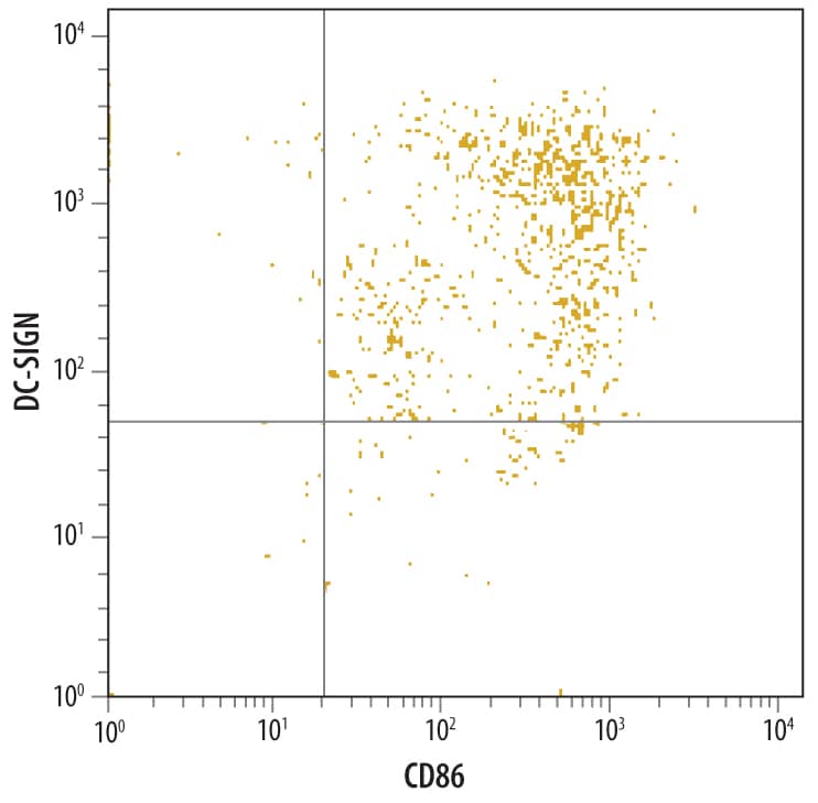
Human monocyte derived dendritic cells were stained with Mouse Anti-Human DC-SIGN Monoclonal Antibody (Catalog # MAB161) followed by PE-conjugated anti-mouse IgG (Catalog # F0102B) and Anti-Human B7-2/CD86 Fluorescein-conjugated Monoclonal Antibody (Catalog # FAB141F). Quadrant markers were set based on control antibody staining (Catalog # MAB0041).Detection of Human DC-SIGN/CD209 by Flow Cytometry
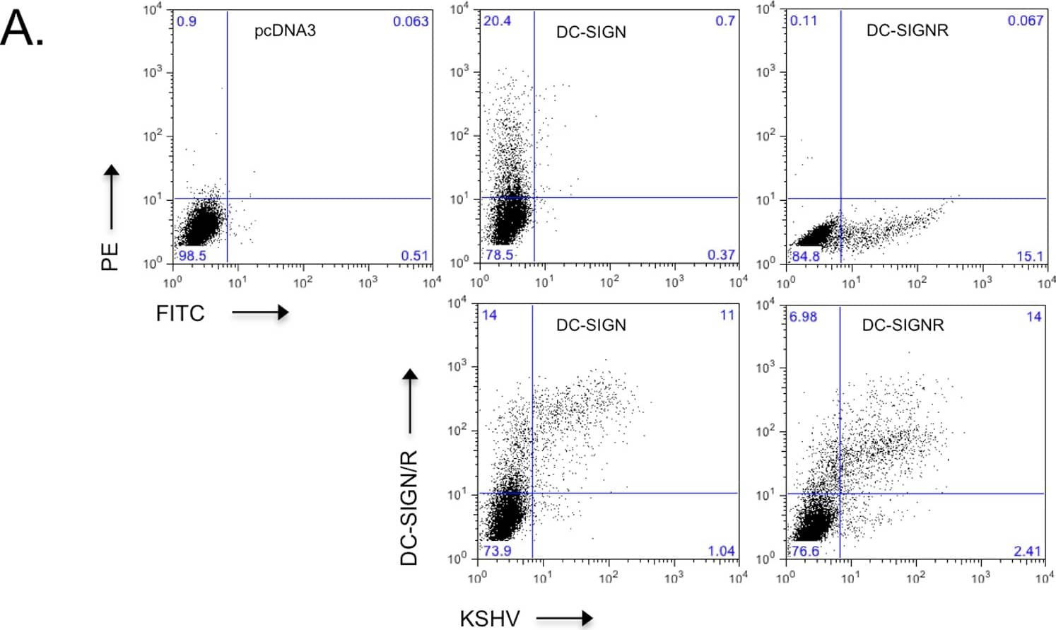
Infectivity of KSHV is enhanced in the presence of DC-SIGN and DC-SIGNR.A) 293T cells were transfected with empty pcDNA3 vector or expression constructs for DC-SIGN or DC-SIGNR. After 24 hours, cells were infected with 20 µl Bac16 deltaK3 deltaK5 or left uninfected as controls. Cells were harvested after additional 24 hours and surface stained with a DC-SIGN/R antibody (H-200) and analyzed by flow cytometry. Top three panels show transfected cells stained for DC-SIGN/R followed by PE- (DC-SIGN), FITC- (DC-SIGNR) or both (vector) conjugated secondary antibodies. Bottom panels shows KSHV infection of 293T cells transiently expressing DC-SIGN or DC-SIGNR. B, left panels) 293 cell lines stably expressing a vector construct, DC-SIGN or DC-SIGNR were fluorescently stained for surface expression of DC-SIGN or DC-SIGNR. The mean channel fluorescence is indicated in the upper right hand corner. Open histograms – secondary antibody alone; shaded histograms – DC-SIGN or DC-SIGNR staining. B, right panel) 293 pcDNA3, DC-SIGN or DC-SIGNR stable cell lines were pre-incubated with a control antibody (anti-ICAM1, 7 µg/ml), with mannan (100 µg/ml) or a monoclonal antibody specific for DC-SIGN (MAB161; 7 µg/ml) for 30 minutes on ice. These cells were then infected with wild type KSHV (Bac16 or rKSHV.219) at an MOI of 0.01. After 72 hours cells were harvested and evaluated for infection by flow cytometry measuring GFP expression. Infection rates were normalized to 293 pcDNA3 cells treated with the control antibody. The fold increase in relative infectivity is indicated. Data are representative of four independent experiments with two performed in triplicate. C) 293 pcDNA3, DC-SIGN or DC-SIGNR stable lines were infected with 50 µl of concentrated Bac16 wildtype (wt), or mutants with deletion of K3 only ( deltaK3), K5 only ( deltaK5), or deletion of both K3 and K5 ( deltaK3 deltaK5) as indicated. At 72 hours post-infection, the cells were stained for surface expression of DC-SIGN, DC-SIGNR or MHC class I. GFP fluorescence was used as a marker for infection. MHC I staining is shown for infected 293 DC-SIGNR cells. Inset numbers indicate percentage of cells in each quadrant. Image collected and cropped by CiteAb from the following publication (https://dx.plos.org/10.1371/journal.pone.0058056), licensed under a CC-BY license. Not internally tested by R&D Systems.Applications for Human DC-SIGN/CD209 Antibody
Adhesion Blockade
CyTOF-ready
Flow Cytometry
Sample: Human DC-SIGN transfected 3T3 mouse embryonic fibroblast cell line and human monocyte derived dendritic cells
Western Blot
Sample: Recombinant Human DC-SIGN Fc Chimera (Catalog # 161-DC)
Reviewed Applications
Read 4 reviews rated 4.3 using MAB161 in the following applications:
Formulation, Preparation, and Storage
Purification
Reconstitution
Formulation
Shipping
Stability & Storage
- 12 months from date of receipt, -20 to -70 °C as supplied.
- 1 month, 2 to 8 °C under sterile conditions after reconstitution.
- 6 months, -20 to -70 °C under sterile conditions after reconstitution.
Background: DC-SIGN/CD209
Human DC-Sign (dendritic cell-specific ICAM-3 grabbing nonintegrin; also CD209) is a member of the chromosome 19 C-type lectin family that includes DC-SIGN, DC-SIGN-related protein, CD23 and LSECtin (1). DC-SIGN was initially reported to be a 46 kDa, 404 amino acid (aa) type II transmembrane protein that contained a 40 aa cytoplasmic N-terminus, a 21 aa transmembrane segment, and a 343 aa extracellular C-terminus (2). The extracellular region contains a distal, 115 aa Ca++-dependent carbohydrate-binding lectin domain and a membrane-proximal linker segment that is composed of seven 23 aa repeats (2, 3). The lectin domain is believed to preferably bind mannose, either within the context of ICAM-3 (on T cells) or ICAM-2 (on endothelial cells) (2, 4, 5). DC-SIGN expression appears to be limited to dendritic cells (DC) and macrophages (6), and DC interaction with the ICAMs both aids DC cell trafficking and immunological synapse formation (7). Since the original report on DC-SIGN, multiple splice forms have been discovered, generating both membrane-bound and soluble forms (3). There are eight type A isoforms, all of which begin with the same 15 aa of exon 1a. Four contain the transmembrane region of exon II, and four do not (i.e., are soluble). Among these eight type A isoforms, only three retain the entire 343 aa found in the full length form described in reference #2 (the full length form is referred to as type I mDC-SIGN1A) (3). Five additional isoforms utilize an alternate start site, and these are referred to as type B isoforms. These all show a 35 aa cytoplasmic domain. One also has a transmembrane segment; four do not. Two of the five contain full, unspliced extracellular regions (3). All of this suggests enormous complexity in DC-SIGN biology. DC-SIGN is not well conserved across species. Human and mouse show little overall aa identity. In the lectin domain, however, human DC-SIGN shares 68% aa identity with mouse DC-SIGN (8). Human and rhesus monkey DC-SIGN share 91% aa identity over the entire extracellular region (8). A detailed description of the additional properties of this monoclonal antibody (MAB161) have been published (9, 10).
References
- Liu, W. et al. (2004) J. Biol. Chem. 279:18748.
- Curtis, B.M. et al. (1992) Proc. Natl. Acad. Sci. USA 89:8356.
- Mummidi, S. et al. (2001) J. Biol. Chem. 276:33196.
- Su, S.V. et al. (2004) J. Biol. Chem. 279:19122.
- Cambi, A. et al. (2005) Cell. Microbiol. 7:481.
- Serrano-Gomez, D. et al. (2004) J. Immunol. 173:5635.
- Geijtenbeek, T.B.H. and Y. van Kooyk (2003) Curr. Top. Microbiol. Immunol. 276:32.
- Baribaud, F. et al. (2001) J. Virol. 75:10281.
- Wu, L. et al. (2002) J. Virol. 76:5905.
- Baribaud, F. et al. (2002) J. Virol.76:9135.
Long Name
Alternate Names
Gene Symbol
Additional DC-SIGN/CD209 Products
Product Documents for Human DC-SIGN/CD209 Antibody
Product Specific Notices for Human DC-SIGN/CD209 Antibody
For research use only


