Human DPPIV/CD26 Antibody
R&D Systems, part of Bio-Techne | Catalog # AF1180

Key Product Details
Species Reactivity
Validated:
Cited:
Applications
Validated:
Cited:
Label
Antibody Source
Product Specifications
Immunogen
Asp34-Pro766
Accession # Q53TN1
Specificity
Clonality
Host
Isotype
Scientific Data Images for Human DPPIV/CD26 Antibody
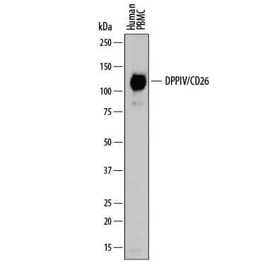
Detection of Human DPPIV/CD26 by Western Blot.
Western blot shows lysate of human peripheral blood mononuclear cells (PBMC). PVDF membrane was probed with 0.2 µg/mL of Goat Anti-Human DPPIV/CD26 Antigen Affinity-purified Polyclonal Antibody (Catalog # AF1180) followed by HRP-conjugated Anti-Goat IgG Secondary Antibody (HAF109). A specific band was detected for DPPIV/CD26 at approximately 110 kDa (as indicated). This experiment was conducted under reducing conditions and using Immunoblot Buffer Group 1.Detection of Human DPPIV/CD26 by Western Blot.
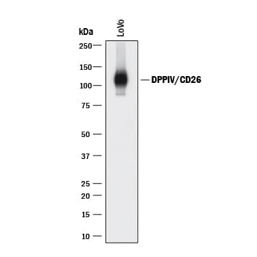
Western blot shows lysate of LoVo human colorectal adenocarcinoma cell line. PVDF membrane was probed with 0.2 µg/mL of Goat Anti-Human DPPIV/CD26 Antigen Affinity-purified Polyclonal Antibody (Catalog # AF1180) followed by HRP-conjugated Anti-Goat IgG Secondary Antibody (HAF017). A specific band was detected for DPPIV/CD26 at approximately 110 kDa (as indicated). This experiment was conducted under reducing conditions and using Immunoblot Buffer Group 1.DPPIV/CD26 in Human PBMCs.
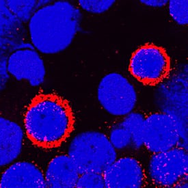
DPPIV/CD26 was detected in immersion fixed human peripheral blood mononuclear cells (PBMCs) using Goat Anti-Human DPPIV/CD26 Antigen Affinity-purified Polyclonal Antibody (Catalog # AF1180) at 1.7 µg/mL for 3 hours at room temperature. Cells were stained using the NorthernLights™ 557-conjugated Anti-Goat IgG Secondary Antibody (red; NL001) and counterstained with DAPI (blue). Specific staining was localized to cytoplasm and plasma membranes. View our protocol for Fluorescent ICC Staining of Non-adherent Cells.Applications for Human DPPIV/CD26 Antibody
Immunocytochemistry
Sample: Immersion fixed human peripheral blood mononuclear cells (PBMCs)
Immunohistochemistry
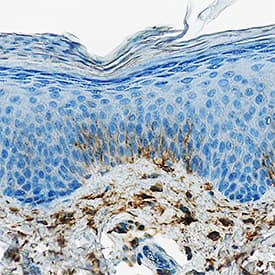
Sample: Immersion fixed paraffin-embedded sections of human prostate and psoriatic skin
Simple Western
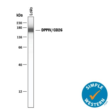
Sample: LoVo human colorectal adenocarcinoma cell line
Western Blot
Sample: Human peripheral blood mononuclear cells (PBMC) and LoVo human colorectal adenocarcinoma cell line
Reviewed Applications
Read 6 reviews rated 4.7 using AF1180 in the following applications:
Formulation, Preparation, and Storage
Purification
Reconstitution
Formulation
Shipping
Stability & Storage
- 12 months from date of receipt, -20 to -70 °C as supplied.
- 1 month, 2 to 8 °C under sterile conditions after reconstitution.
- 6 months, -20 to -70 °C under sterile conditions after reconstitution.
Background: DPPIV/CD26
DPPIV/CD26 plays an important role in many biological and pathological processes. It functions as T cell-activating molecule (THAM). It serves as a cofactor for entry of HIV in CD4+ cells. It binds adenosine deaminase, the deficiency of which causes severe combined immunodeficiency disease in humans. It cleaves chemokines such as stromal-cell-derived factor 1 alpha and macrophage-derived chemokine. It degrades peptide hormones such as glucagon. It truncates procalcitonin, a marker for systemic bacterial infections with elevated levels detected in patients with thermal injury, sepsis and severe infection, and in children with bacterial meningitis.
Long Name
Alternate Names
Gene Symbol
UniProt
Additional DPPIV/CD26 Products
Product Documents for Human DPPIV/CD26 Antibody
Product Specific Notices for Human DPPIV/CD26 Antibody
For research use only
