Human Drebrin 1 Antibody
R&D Systems, part of Bio-Techne | Catalog # MAB7739

Key Product Details
Species Reactivity
Human
Applications
Immunocytochemistry, Simple Western, Western Blot
Label
Unconjugated
Antibody Source
Monoclonal Mouse IgG1 Clone # 838102
Product Specifications
Immunogen
E. coli-derived recombinant human Drebrin 1
Asn482-Asp649 (Ser553Pro)
Accession # Q16643
Asn482-Asp649 (Ser553Pro)
Accession # Q16643
Specificity
Detects human Drebrin 1 in ELISAs.
Clonality
Monoclonal
Host
Mouse
Isotype
IgG1
Scientific Data Images for Human Drebrin 1 Antibody
Detection of Human Drebrin 1 by Western Blot.
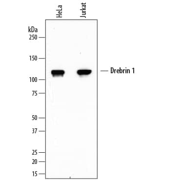
Western blot shows lysates of HeLa human cervical epithelial carcinoma cell line and Jurkat human acute T cell leukemia cell line. PVDF membrane was probed with 1 µg/mL of Mouse Anti-Human Drebrin 1 Monoclonal Antibody (Catalog # MAB7739) followed by HRP-conjugated Anti-Mouse IgG Secondary Antibody (Catalog # HAF007). A specific band was detected for Drebrin 1 at approximately 120 kDa (as indicated). This experiment was conducted under reducing conditions and using Immunoblot Buffer Group 1.Drebrin 1 in HeLa Human Cell Line.
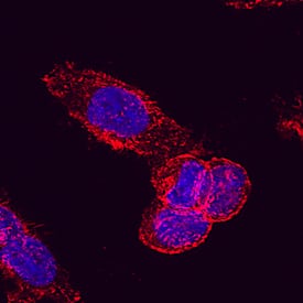
Drebrin 1 was detected in immersion fixed HeLa human cervical epithelial carcinoma cell line using Mouse Anti-Human Drebrin 1 Monoclonal Antibody (Catalog # MAB7739) at 25 µg/mL for 3 hours at room temperature. Cells were stained using the Northern-Lights™ 557-conjugated Anti-Mouse IgG Secondary Antibody (red; Catalog # NL007) and counterstained with DAPI (blue). Specific staining was localized to cell membrane and cytoplasm. View our protocol for Fluorescent ICC Staining of Cells on Coverslips.Detection of Human Drebrin 1 by Simple WesternTM.
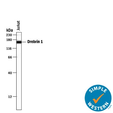
Simple Western lane view shows lysates of Jurkat human acute T cell leukemia cell line, loaded at 0.5 mg/mL. A specific band was detected for Drebrin 1 at approximately 162 kDa (as indicated) using 10 µg/mL of Mouse Anti-Human Drebrin 1 Monoclonal Antibody (Catalog # MAB7739) . This experiment was conducted under reducing conditions and using the 12-230 kDa separation system.Applications for Human Drebrin 1 Antibody
Application
Recommended Usage
Immunocytochemistry
8-25 µg/mL
Sample: Immersion fixed HeLa human cervical epithelial carcinoma cell line
Sample: Immersion fixed HeLa human cervical epithelial carcinoma cell line
Simple Western
10 µg/mL
Sample: Jurkat human acute T cell leukemia cell line
Sample: Jurkat human acute T cell leukemia cell line
Western Blot
1 µg/mL
Sample: HeLa human cervical epithelial carcinoma cell line and Jurkat human acute T cell leukemia cell line
Sample: HeLa human cervical epithelial carcinoma cell line and Jurkat human acute T cell leukemia cell line
Reviewed Applications
Read 1 review rated 4 using MAB7739 in the following applications:
Formulation, Preparation, and Storage
Purification
Protein A or G purified from hybridoma culture supernatant
Reconstitution
Sterile PBS to a final concentration of 0.5 mg/mL. For liquid material, refer to CoA for concentration.
Formulation
Lyophilized from a 0.2 μm filtered solution in PBS with Trehalose. *Small pack size (SP) is supplied either lyophilized or as a 0.2 µm filtered solution in PBS.
Shipping
Lyophilized product is shipped at ambient temperature. Liquid small pack size (-SP) is shipped with polar packs. Upon receipt, store immediately at the temperature recommended below.
Stability & Storage
Use a manual defrost freezer and avoid repeated freeze-thaw cycles.
- 12 months from date of receipt, -20 to -70 °C as supplied.
- 1 month, 2 to 8 °C under sterile conditions after reconstitution.
- 6 months, -20 to -70 °C under sterile conditions after reconstitution.
Background: Drebrin 1
Alternate Names
D0S117E, DBN1, Drba, Drebrin A, Drebrin E, Drebrin E2
Gene Symbol
DBN1
UniProt
Additional Drebrin 1 Products
Product Documents for Human Drebrin 1 Antibody
Product Specific Notices for Human Drebrin 1 Antibody
For research use only
Loading...
Loading...
Loading...
Loading...


