Human ER alpha/NR3A1 Antibody
R&D Systems, part of Bio-Techne | Catalog # AF5715

Key Product Details
Species Reactivity
Validated:
Human
Cited:
Human
Applications
Validated:
Immunohistochemistry, Simple Western, Western Blot
Cited:
Functional Assay
Label
Unconjugated
Antibody Source
Polyclonal Sheep IgG
Product Specifications
Immunogen
E. coli-derived recombinant human ER alpha
Met1-Gln116
Accession # P03372
Met1-Gln116
Accession # P03372
Specificity
Detects human ER alpha.
Clonality
Polyclonal
Host
Sheep
Isotype
IgG
Scientific Data Images for Human ER alpha/NR3A1 Antibody
ER alpha/NR3A1 in Human Endometrial Cancer Tissue.
ERa/NR3A1 was detected in immersion fixed paraffin-embedded sections of human endometrial cancer tissue using Sheep Anti-Human ERa/NR3A1 Antigen Affinity-purified Polyclonal Antibody (Catalog # AF5715) at 10 µg/mL overnight at 4 °C. Before incubation with the primary antibody tissue was subjected to heat-induced epitope retrieval using Antigen Retrieval Reagent-Basic (Catalog # CTS013). Tissue was stained using the Anti-Sheep HRP-DAB Cell & Tissue Staining Kit (brown; Catalog # CTS019) and counterstained with hematoxylin (blue). View our protocol for Chromogenic IHC Staining of Paraffin-embedded Tissue Sections.Detection of Human ER alpha/NR3A1 by Western Blot.
Western blot shows lysates of MCF-7 human breast cancer cell line and MDA-MB-468 human breast cancer cell line. PVDF membrane was probed with 0.5 µg/mL of Sheep Anti-Human ERa/NR3A1 Antigen Affinity-purified Polyclonal Antibody (Catalog # AF5715) followed by HRP-conjugated Anti-Sheep IgG Secondary Antibody (Catalog # HAF016). A specific band was detected for ERa/NR3A1 at approximately 65 to 70 kDa (as indicated). This experiment was conducted under reducing conditions and using Immunoblot Buffer Group 1.Detection of Human ER alpha/NR3A1 by Simple WesternTM.
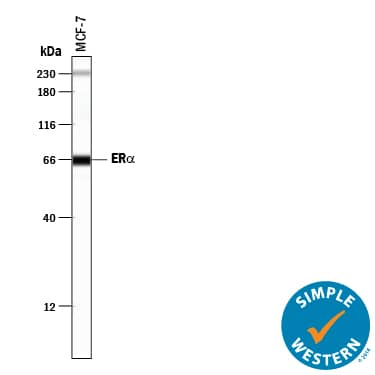
Simple Western lane view shows lysates of MCF-7 human breast cancer cell line, loaded at 0.2 mg/mL. A specific band was detected for ERa/NR3A1 at approximately 66 kDa (as indicated) using 5 µg/mL of Sheep Anti-Human ERa/NR3A1 Antigen Affinity-purified Polyclonal Antibody (Catalog # AF5715) followed by 1:50 dilution of HRP-conjugated Anti-Sheep IgG Secondary Antibody (Catalog # HAF016). This experiment was conducted under reducing conditions and using the 12-230 kDa separation system. Non-specific interaction with the 230 kDa Simple Western standard may be seen with this antibody.Applications for Human ER alpha/NR3A1 Antibody
Application
Recommended Usage
Immunohistochemistry
5-15 µg/mL
Sample: Immersion fixed paraffin-embedded sections of human endometrial cancer tissue subjected to Antigen Retrieval Reagent-Basic (Catalog # CTS013)
Sample: Immersion fixed paraffin-embedded sections of human endometrial cancer tissue subjected to Antigen Retrieval Reagent-Basic (Catalog # CTS013)
Simple Western
5 µg/mL
Sample: MCF‑7 human breast cancer cell line
Sample: MCF‑7 human breast cancer cell line
Western Blot
0.5 µg/mL
Sample: MCF-7 human breast cancer cell line and MDA-MB-468 human breast cancer cell line
Sample: MCF-7 human breast cancer cell line and MDA-MB-468 human breast cancer cell line
Formulation, Preparation, and Storage
Purification
Antigen Affinity-purified
Reconstitution
Reconstitute at 0.2 mg/mL in sterile PBS. For liquid material, refer to CoA for concentration.
Formulation
Lyophilized from a 0.2 μm filtered solution in PBS with Trehalose. *Small pack size (SP) is supplied either lyophilized or as a 0.2 µm filtered solution in PBS.
Shipping
Lyophilized product is shipped at ambient temperature. Liquid small pack size (-SP) is shipped with polar packs. Upon receipt, store immediately at the temperature recommended below.
Stability & Storage
Use a manual defrost freezer and avoid repeated freeze-thaw cycles.
- 12 months from date of receipt, -20 to -70 °C as supplied.
- 1 month, 2 to 8 °C under sterile conditions after reconstitution.
- 6 months, -20 to -70 °C under sterile conditions after reconstitution.
Background: ER alpha/NR3A1
Long Name
Estrogen Receptor alpha
Alternate Names
ESR1, NR3A1
Entrez Gene IDs
2099 (Human)
Gene Symbol
ESR1
UniProt
Additional ER alpha/NR3A1 Products
Product Documents for Human ER alpha/NR3A1 Antibody
Product Specific Notices for Human ER alpha/NR3A1 Antibody
For research use only
Loading...
Loading...
Loading...
Loading...


