Human Fc gamma RIII (CD16) Antibody
R&D Systems, part of Bio-Techne | Catalog # MAB2546


Conjugate
Catalog #
Key Product Details
Species Reactivity
Validated:
Human
Cited:
Human
Applications
Validated:
CyTOF-ready, Flow Cytometry
Cited:
Flow Cytometry, Functional Assay, Immunocytochemistry, Immunoprecipitation
Label
Unconjugated
Antibody Source
Monoclonal Mouse IgG2A Clone # 245536
Product Specifications
Immunogen
Mouse myeloma cell line NS0-derived recombinant human Fc gamma RIIIA/B (CD16)
Thr20-Gln208
Accession # O75015
Thr20-Gln208
Accession # O75015
Specificity
Detects human Fc gamma RIIIA/B (CD16) in direct ELISAs. In direct ELISAs, approximately 10% cross-reactivity with recombinant human Fc gamma RIIA and recombinant mouse Fc gamma RIII is observed.
Clonality
Monoclonal
Host
Mouse
Isotype
IgG2A
Scientific Data Images for Human Fc gamma RIII (CD16) Antibody
Detection of Fc gamma RIIIA/B (CD16a/b) in Human PBMCs by Flow Cytometry.
Human peripheral blood mononuclear cells (PBMCs) were stained with (A) Mouse Anti-Human Fc gamma RIII A/B (CD16a/b) Monoclonal Antibody (Catalog # MAB2546) or (B) Mouse IgG2A isotype control antibody (MAB003) followed by Goat anti-Mouse IgG APC-conjugated Secondary Antibody (F0101B) and Mouse Anti-Human CD14 PE-conjugated Monoclonal Antibody (FAB3832P). Staining was performed using our Staining Membrane-associated Proteins protocol.Detection of Human Fc gamma RIII (CD16) by Functional
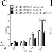
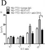
Anti-ULBP3 monoclonal antibody enhances the activity of NK cells from cancer patients via ADCC.ULBP3 expression and the percentage of infiltrating NK cells in tumor tissues from 10 patients (4 colorectal cancer, 3 lung cancer, and 3 gastric cancer) were determined by FCM. A representative graph is shown in (A). The relationship between ULBP3 expression and infiltrating NK cells in tumor tissues is shown in (B). Normal NK cells were co-cultured with CD133−SW620 (C) or 7721 (D) cells at the indicated E:T ratios, and cytotoxicity was measured by LDH release after 6 h of co-culture, with or without the addition of isotype IgG, anti-ULBP3 (B2-F1-F1), and/or anti-CD16 (5 μg/ml). K562 (E) or A549 (F) cells were co-cultured with normal NK cells, and cytotoxicity was measured by LDH release after 6 h of co-culture at the indicated E:T ratios, with or without the addition of ULBP3-Fc (1 ng/ml) and/or B2-F1-F1 (5 μg/ml). The data are representative of results obtained from at least 3 independent experiments. The results are expressed as the mean ± SEM. *P < 0.05, **P < 0.01, ***P < 0.001. Image collected and cropped by CiteAb from the following publication (https://www.nature.com/articles/srep06138), licensed under a CC-BY license. Not internally tested by R&D Systems.Detection of Human Fc gamma RIII (CD16) by Functional
Anti-ULBP3 monoclonal antibody enhances the activity of NK cells from cancer patients via ADCC.ULBP3 expression and the percentage of infiltrating NK cells in tumor tissues from 10 patients (4 colorectal cancer, 3 lung cancer, and 3 gastric cancer) were determined by FCM. A representative graph is shown in (A). The relationship between ULBP3 expression and infiltrating NK cells in tumor tissues is shown in (B). Normal NK cells were co-cultured with CD133−SW620 (C) or 7721 (D) cells at the indicated E:T ratios, and cytotoxicity was measured by LDH release after 6 h of co-culture, with or without the addition of isotype IgG, anti-ULBP3 (B2-F1-F1), and/or anti-CD16 (5 μg/ml). K562 (E) or A549 (F) cells were co-cultured with normal NK cells, and cytotoxicity was measured by LDH release after 6 h of co-culture at the indicated E:T ratios, with or without the addition of ULBP3-Fc (1 ng/ml) and/or B2-F1-F1 (5 μg/ml). The data are representative of results obtained from at least 3 independent experiments. The results are expressed as the mean ± SEM. *P < 0.05, **P < 0.01, ***P < 0.001. Image collected and cropped by CiteAb from the following publication (https://www.nature.com/articles/srep06138), licensed under a CC-BY license. Not internally tested by R&D Systems.Applications for Human Fc gamma RIII (CD16) Antibody
Application
Recommended Usage
CyTOF-ready
Ready to be labeled using established conjugation methods. No BSA or other carrier proteins that could interfere with conjugation.
Flow Cytometry
0.25 µg/106 cells
Sample: Human PBMC
Sample: Human PBMC
Formulation, Preparation, and Storage
Purification
Protein A or G purified from hybridoma culture supernatant
Reconstitution
Reconstitute at 0.5 mg/mL in sterile PBS. For liquid material, refer to CoA for concentration.
Formulation
Lyophilized from a 0.2 μm filtered solution in PBS with Trehalose. *Small pack size (SP) is supplied either lyophilized or as a 0.2 µm filtered solution in PBS.
Shipping
Lyophilized product is shipped at ambient temperature. Liquid small pack size (-SP) is shipped with polar packs. Upon receipt, store immediately at the temperature recommended below.
Stability & Storage
Use a manual defrost freezer and avoid repeated freeze-thaw cycles.
- 12 months from date of receipt, -20 to -70 °C as supplied.
- 1 month, 2 to 8 °C under sterile conditions after reconstitution.
- 6 months, -20 to -70 °C under sterile conditions after reconstitution.
Background: Fc gamma RIII (CD16)
Long Name
Fc gamma Receptor III
Alternate Names
FcgRIII
Gene Symbol
FCGR3
UniProt
Additional Fc gamma RIII (CD16) Products
Product Documents for Human Fc gamma RIII (CD16) Antibody
Product Specific Notices for Human Fc gamma RIII (CD16) Antibody
For research use only
Loading...
Loading...
Loading...
Loading...
Loading...

