Human GILT/IFI30 Antibody
R&D Systems, part of Bio-Techne | Catalog # MAB7715

Key Product Details
Species Reactivity
Human
Applications
Immunocytochemistry, Western Blot
Label
Unconjugated
Antibody Source
Monoclonal Mouse IgG2A Clone # 844623
Product Specifications
Immunogen
HEK293 human embryonic kidney cell line transfected with human GILT/IFI30
Ser27-Lys250
Accession # P13284
Ser27-Lys250
Accession # P13284
Specificity
Detects human GILT/IFI30 in direct ELISAs and Western blots.
Clonality
Monoclonal
Host
Mouse
Isotype
IgG2A
Scientific Data Images for Human GILT/IFI30 Antibody
Detection of Human GILT/IFI30 by Western Blot.
Western blot shows lysates of Raji human Burkitt's lymphoma cell line. PVDF membrane was probed with 1 µg/mL of Mouse Anti-Human GILT/IFI30 Monoclonal Antibody (Catalog # MAB7715) followed by HRP-conjugated Anti-Mouse IgG Secondary Antibody (Catalog # HAF018). Specific bands were detected for GILT/IFI30 at approximately 25-30 kDa (as indicated). This experiment was conducted under reducing conditions and using Immunoblot Buffer Group 1.GILT/IFI30 in THP‑1 Human Cell Line.
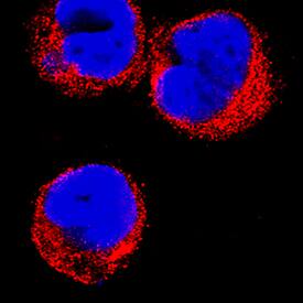
GILT/IFI30 was detected in immersion fixed THP-1 human acute monocytic leukemia cell line using Mouse Anti-Human GILT/IFI30 Monoclonal Antibody (Catalog # MAB7715) at 8 µg/mL for 3 hours at room temperature. Cells were stained using the NorthernLights™ 557-conjugated Anti-Mouse IgG Secondary Antibody (red; Catalog # NL007) and counterstained with DAPI (blue). Specific staining was localized to cytoplasm. View our protocol for Fluorescent ICC Staining of Non-adherent Cells.GILT/IFI30 in MCF‑7 Human Cell Line.
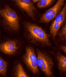
GILT/IFI30 was detected in immersion fixed MCF-7 human breast cancer cell line using Mouse Anti-Human GILT/IFI30 Monoclonal Antibody (Catalog # MAB7715) at 10 µg/mL for 3 hours at room temperature. Cells were stained using the NorthernLights™ 557-conjugated Anti-Mouse IgG Secondary Antibody (red; Catalog # NL007) and counterstained with DAPI (blue). Specific staining was localized to cytoplasm. View our protocol for Fluorescent ICC Staining of Cells on Coverslips.Applications for Human GILT/IFI30 Antibody
Application
Recommended Usage
Immunocytochemistry
5-25 µg/mL
Sample: Immersion fixed THP‑1 human acute monocytic leukemia cell line and MCF-7 human breast cancer cell line
Sample: Immersion fixed THP‑1 human acute monocytic leukemia cell line and MCF-7 human breast cancer cell line
Western Blot
1 µg/mL
Sample: Raji human Burkitt's lymphoma cell line
Sample: Raji human Burkitt's lymphoma cell line
Formulation, Preparation, and Storage
Purification
Protein A or G purified from hybridoma culture supernatant
Reconstitution
Sterile PBS to a final concentration of 0.5 mg/mL. For liquid material, refer to CoA for concentration.
Formulation
Lyophilized from a 0.2 μm filtered solution in PBS with Trehalose. *Small pack size (SP) is supplied either lyophilized or as a 0.2 µm filtered solution in PBS.
Shipping
Lyophilized product is shipped at ambient temperature. Liquid small pack size (-SP) is shipped with polar packs. Upon receipt, store immediately at the temperature recommended below.
Stability & Storage
Use a manual defrost freezer and avoid repeated freeze-thaw cycles.
- 12 months from date of receipt, -20 to -70 °C as supplied.
- 1 month, 2 to 8 °C under sterile conditions after reconstitution.
- 6 months, -20 to -70 °C under sterile conditions after reconstitution.
Background: GILT/IFI30
N‑ and C-terminus, a 175 aa, 25-30 kDa active mature form is generated (aa 58-232). The mature region possesses a thiol reductase domain (aa 62-151) plus one utilized Thr phosphorylation site. Both the pro‑ and mature forms exhibit enzymatic activity. IFI30 is known to exist as a 50-60 kDa disulfide-linked homodimer. There are four potential isoform variants. One contains a 26 aa substitution for aa 213-250, a second shows a deletion of aa 131-161, a third shows a deletion of aa 106-123, while a fourth shows a deletion of aa 64-212. Over aa 27-250, human IFI30 shares 62% aa sequence identity with mouse IFI30.
Long Name
Gamma-Interferon-inducible Lysosomal Thiol Reductase
Alternate Names
IFI30, IP30, Legumaturain
Gene Symbol
IFI30
UniProt
Additional GILT/IFI30 Products
Product Documents for Human GILT/IFI30 Antibody
Product Specific Notices for Human GILT/IFI30 Antibody
For research use only
Loading...
Loading...
Loading...
Loading...


