Human GPR56 Antibody
R&D Systems, part of Bio-Techne | Catalog # MAB46361


Key Product Details
Species Reactivity
Applications
Label
Antibody Source
Product Specifications
Immunogen
Met1-Val345
Accession # Q9Y653
Specificity
Clonality
Host
Isotype
Scientific Data Images for Human GPR56 Antibody
Detection of Human GPR56 by Western Blot.
Western blot shows lysates of NS0 mouse myeloma cell line either mock transfected or transfected with human GPR56. PVDF membrane was probed with 2 µg/mL of Rabbit Anti-Human GPR56 Monoclonal Antibody (Catalog # MAB46361) followed by HRP-conjugated Anti-Rabbit IgG Secondary Antibody (Catalog # HAF008). A specific band was detected for GPR56 at approximately 75 kDa (as indicated). GAPDH (Catalog # AF5718) is shown as a loading control.This experiment was conducted under reducing conditions and using Immunoblot Buffer Group 1.Detection of GPR56 in Human Peripheral Blood Cells by Flow Cytometry.
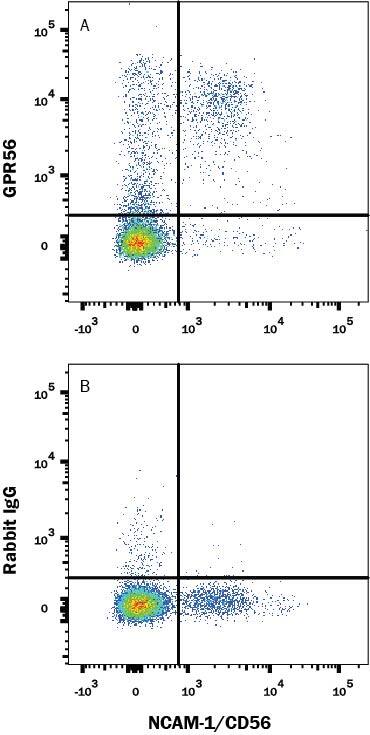
Human peripheral blood cells were stained with (A) Rabbit Anti-Human GPR56 Monoclonal Antibody (Catalog # MAB46361) or (B) Rabbit IgG isotype control antibody (Catalog # MAB1050) followed by APC-conjugated Anti-Rabbit IgG Secondary Antibody (Catalog # F0111) and Mouse anti-Human CD56 PE-conjugated monoclonal antibody (Catalog # FAB2408P). View our protocol for Staining Membrane-associated Proteins.GPR56 in T47D human breast cancer cell line.
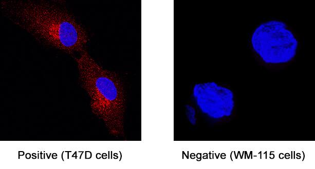
GPR56 was detected in immersion fixed T47D human breast cancer cell line (left panel; positive staining) and WM-115 human malignant melanoma cell line (right panel; negative staining) using Rabbit Anti-Human GPR56 Monoclonal Antibody (Catalog # MAB46361) at 3 µg/mL for 3 hours at room temperature. Cells were stained using the NorthernLights™ 557-conjugated Anti-Rabbit IgG Secondary Antibody (red; Catalog # NL004) and counterstained with DAPI (blue). Specific staining was localized to cytoplasm. View our protocol for Fluorescent ICC Staining of Cells on Coverslips.Applications for Human GPR56 Antibody
CyTOF-ready
Flow Cytometry
Sample: Human Peripheral Blood Cells
Immunocytochemistry
Sample: Immersion fixed T47D human breast cancer cell line
Western Blot
Sample: NS0 mouse myeloma cell line transfected with human GPR56
Reviewed Applications
Read 1 review rated 5 using MAB46361 in the following applications:
Formulation, Preparation, and Storage
Purification
Reconstitution
Formulation
Shipping
Stability & Storage
- 12 months from date of receipt, -20 to -70 °C as supplied.
- 1 month, 2 to 8 °C under sterile conditions after reconstitution.
- 6 months, -20 to -70 °C under sterile conditions after reconstitution.
Background: GPR56
GPR56 is a member of the LN-TM7 family of adhesion-type 7-transmembrane (TM) G-protein coupled receptors (GPCR) with long extracellular N-termini (1‑3). The 693 amino acid (aa) human GPR56 contains a 25 aa signal sequence, a 377 aa N-terminal extracellular domain (ECD) and seven TM regions separated by short intracellular and extracellular regions. Like other LN-TM7 members, the ECD contains a highly glycosylated mucin-like stalk followed by a GPCR proteolytic cleavage site (GPS) (1, 4). Cleavage of the 60 kDa N-terminus from the 80 kDa full length form is needed for efficient cell surface expression (5, 6). While the cleaved portion may remain non-covalently associated, it has also been found in conditioned medium of cultured cells (5). Human GPR56 shares 71%, 72%, 80%, 80% and 79% aa identity with mouse, rat, canine, equine, and bovine GPR56 within the cleaved ECD. A functional splice variant lacking the GPS site and a non-functional splice variant lacking portions of the TM domains have also been described (4). A human brain developmental disorder, bilateral frontoparietal polymicrogyria, is associated with GPR56 mutations that also show impaired GPS cleavage, intracellular trafficking, and expression at the cell surface (5). GPR56 is widely distributed, with highest mRNA or expressed sequence tag expression in brain, thyroid, skin and female reproductive system (3, 4). GPR56 expression is upregulated during cell transformation and is high in melanomas, glioblastomas and astrocytomas, but downregulated in melanomas with high metastatic potential (2, 6‑8). Although the function of GPR56 is not completely known, it is clearly an adhesion protein (6‑8). Tissue transglutaminase (TG2) is one reported ligand, binding of which inhibits melanoma growth and metastasis (6). Association of GPR56 with the tetraspanin CD81 stabilizes its complex with Gaq/11 for cell signaling (9).
References
- Fredriksson, R. et al. (2002) FEBS Lett. 531:407.
- Zendman, A.J.W. et al. (1999) FEBS Lett. 446:292.
- Liu, M. et al. (1999) Genomics 55:296.
- Bjarnadottir, T.K. et al. (2007) Gene 387:38.
- Jin, Z. et al. (2007) Hum. Mol. Genet. 16:1972.
- Xu, L. et al. (2006) Proc. Natl. Acad. Sci. USA 103:9023.
- Shashidhar, S. et al. (2005) Oncogene 24:1673.
- Ke, N. et al. (2007) Mol. Cancer Ther. 6:1840.
- Little, K.D. et al. (2004) Mol. Biol. Cell 15:2375.
Long Name
Alternate Names
Gene Symbol
UniProt
Additional GPR56 Products
Product Documents for Human GPR56 Antibody
Product Specific Notices for Human GPR56 Antibody
For research use only

