Human ICAP-1 Antibody
R&D Systems, part of Bio-Techne | Catalog # AF6805

Key Product Details
Species Reactivity
Validated:
Cited:
Applications
Validated:
Cited:
Label
Antibody Source
Product Specifications
Immunogen
Met1-Tyr127
Accession # O14713
Specificity
Clonality
Host
Isotype
Scientific Data Images for Human ICAP-1 Antibody
Detection of Human ICAP‑1 by Western Blot.
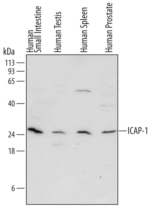
Western blot shows lysates of human small intestine tissue, human testis tissue, human spleen tissue, and human prostate tissue. PVDF Membrane was probed with 2 µg/mL of Sheep Anti-Human ICAP-1 Antigen Affinity-purified Polyclonal Antibody (Catalog # AF6805) followed by HRP-conjugated Anti-Sheep IgG Secondary Antibody (Catalog # HAF016). A specific band was detected for ICAP-1 at approximately 25 kDa (as indicated). This experiment was conducted under reducing conditions and using Immunoblot Buffer Group 1.ICAP-1 in Human Colon Cancer Tissue.
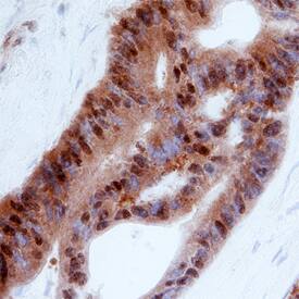
ICAP-1 was detected in immersion fixed paraffin-embedded sections of human colon cancer tissue using Sheep Anti-Human ICAP-1 Antigen Affinity-purified Polyclonal Antibody (Catalog # AF6805) at 3 µg/mL overnight at 4 °C. Before incubation with the primary antibody, tissue was subjected to heat-induced epitope retrieval using Antigen Retrieval Reagent-Basic (Catalog # CTS013). Tissue was stained using the Anti-Sheep HRP-DAB Cell & Tissue Staining Kit (brown; Catalog # CTS019) and counterstained with hematoxylin (blue). Specific staining was localized to nuclei in epithelial cells. View our protocol for Chromogenic IHC Staining of Paraffin-embedded Tissue Sections.Applications for Human ICAP-1 Antibody
Immunohistochemistry
Sample: Immersion fixed paraffin-embedded sections of human colon cancer tissue
Western Blot
Sample: Human small intestine tissue, human testis tissue, human spleen tissue, and human prostate tissue
Formulation, Preparation, and Storage
Purification
Reconstitution
Formulation
Shipping
Stability & Storage
- 12 months from date of receipt, -20 to -70 °C as supplied.
- 1 month, 2 to 8 °C under sterile conditions after reconstitution.
- 6 months, -20 to -70 °C under sterile conditions after reconstitution.
Background: ICAP-1
ICAP-1 (Integrin cytoplasmic domain-associated protein-1; also ITBP1) is a 22-27 kDa protein that specifically binds to the cytoplasmic tail of beta1 integrin. It is widely expressed, and serves to negatively regulate cell migration/proliferation. In the cytosol, it has at least two functions. First, it associates with beta1 integrin, promoting an inactive conformation. This has the effect of both slowing cell migration and allowing for the monitoring of ECM density. Second, it induces Delta-Notch signaling, an event that specifies the growth of only tip endothelium during angiogenesis. In the nucleus, ICAP-1 is proposed to participate in c-myc activation. Human ICAP-1 is 200 amino acids (aa) in length. It contains an NLS (aa 5-9) and one pleckstrin homology-like/PID domain (aa 58-200). Phosphorylation occurs at Thr38 and Ser41. There is one nonintegrin-binding alternative splice form that shows a deletion of aa 128-177. Over aa 1-127, human ICAP-1 shares 93% aa identity with mouse ICAP-1.
Long Name
Alternate Names
Gene Symbol
UniProt
Additional ICAP-1 Products
Product Documents for Human ICAP-1 Antibody
Product Specific Notices for Human ICAP-1 Antibody
For research use only

