Human IFN-alpha / beta R1 Antibody
R&D Systems, part of Bio-Techne | Catalog # MAB2452
Recombinant Monoclonal Antibody.


Conjugate
Catalog #
Key Product Details
Species Reactivity
Human
Applications
Flow Cytometry, Western Blot
Label
Unconjugated
Antibody Source
Recombinant Monoclonal Rabbit IgG Clone # 2951C
Product Specifications
Immunogen
Mouse myeloma cell line, NS0-derived human IFN-alpha/beta R1
Gly26-Lys436
Accession # P17181
Gly26-Lys436
Accession # P17181
Specificity
Detects human IFN‑ alpha/ beta R1 in direct ELISA.
Clonality
Monoclonal
Host
Rabbit
Isotype
IgG
Scientific Data Images for Human IFN-alpha / beta R1 Antibody
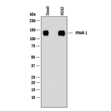
Detection of Human IFN-alpha / beta R1 by Western Blot.
Western blot shows lysates of Daudi human Burkitt's lymphoma cell line and K562 human chronic myelogenous leukemia cell line. PVDF membrane was probed with 2 µg/mL of Rabbit Anti-Human IFN-alpha / beta R1 Monoclonal Antibody (Catalog # MAB2452) followed by HRP-conjugated Anti-Rabbit IgG Secondary Antibody (Catalog # HAF008). A specific band was detected for IFN-alpha / beta R1 at approximately 120 kDa (as indicated). This experiment was conducted under reducing conditions and using Western Blot Buffer Group 1.Detection of IFN-alpha / beta R1 in U937 cells by Flow Cytometry.
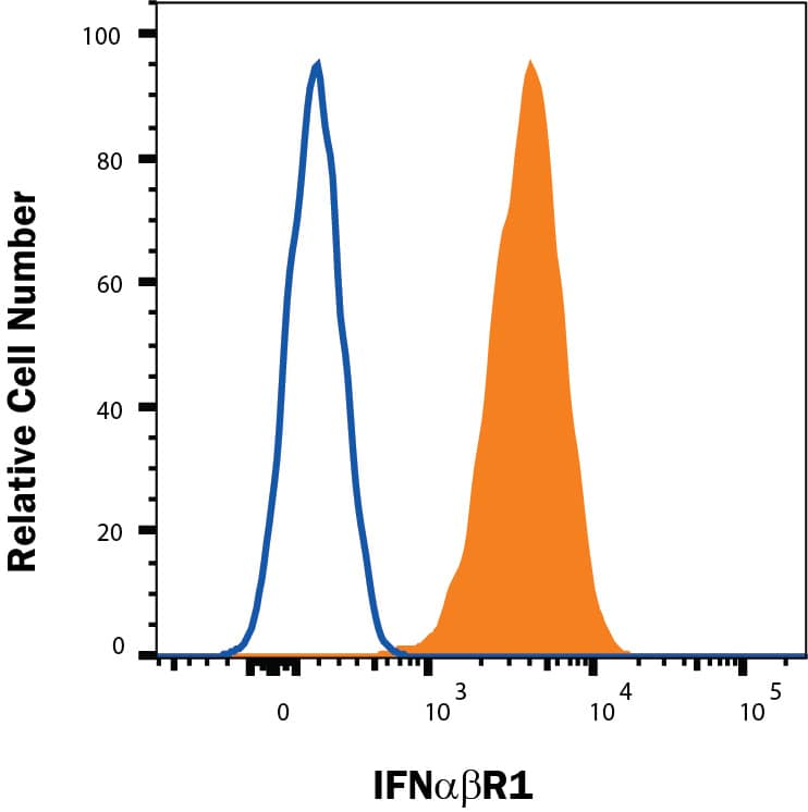
U937 cells were stained with Rabbit Anti-Human IFN-alpha / beta R1 Monoclonal Antibody (Catalog # MAB2452, filled histogram) or isotype control antibody (Catalog # MAB1050, open histogram), followed by Allophycocyanin-conjugated Anti-Rabbit IgG Secondary Antibody (Catalog # F0111). View our protocol for Staining Membrane-associated Proteins.Applications for Human IFN-alpha / beta R1 Antibody
Application
Recommended Usage
Flow Cytometry
0.10 µg/106 cells
Sample: U937 human histiocytic lymphoma cell line
Sample: U937 human histiocytic lymphoma cell line
Western Blot
2 µg/mL
Sample: Daudi human Burkitt's lymphoma cell line and K562 human chronic myelogenous leukemia cell line
Sample: Daudi human Burkitt's lymphoma cell line and K562 human chronic myelogenous leukemia cell line
Formulation, Preparation, and Storage
Purification
Protein A or G purified from hybridoma culture supernatant
Reconstitution
Reconstitute at 0.5 mg/mL in sterile PBS. For liquid material, refer to CoA for concentration.
Formulation
Lyophilized from a 0.2 μm filtered solution in PBS with Trehalose. *Small pack size (SP) is supplied either lyophilized or as a 0.2 µm filtered solution in PBS.
Shipping
Lyophilized product is shipped at ambient temperature. Liquid small pack size (-SP) is shipped with polar packs. Upon receipt, store immediately at the temperature recommended below.
Stability & Storage
Use a manual defrost freezer and avoid repeated freeze-thaw cycles.
- 12 months from date of receipt, -20 to -70 °C as supplied.
- 1 month, 2 to 8 °C under sterile conditions after reconstitution.
- 6 months, -20 to -70 °C under sterile conditions after reconstitution.
Background: IFN-alpha/beta R1
References
- Langer, J.A. et al. (2004) Cytokine Growth Factor Rev. 15:33.
- Hwang, S.Y. et al. (1995) Proc. Natl. Acad. Sci. USA 92:11284.
- Takaoka, A. et al. (2000) Science 288:2357.
- Uze, G. et al. (1990) Cell 60:225.
- Lamken, P. et al. (2004) J. Mol. Biol. 341:303.
- Arduini, R.M. et al. (1999) Prot. Sci. 8:1867.
- Kalie, E. et al. (2008) J. Biol. Chem. 283:32925.
- Platanias, L.C. (2005) Nat. Rev. Immunol. 5:375.
- Claudinon, J. et al. (2009) J. Biol. Chem. 284:24328.
- Zheng, H. et al. (2011) Blood 118:4003.
- Qian, J. et al. (2011) PLoS Pathogens 7:e1002065.
- Bhattacharya, S. et al. (2010) J. Biol. Chem. 285:2318.
- Bhattacharya, S. et al. (2011) J. Biol. Chem. 286:22069.
Long Name
Interferon alpha/beta Receptor 1
Alternate Names
IFN-aR1, IFN-bR1, IFNAR1, IFNbR1
Gene Symbol
IFNAR1
UniProt
Additional IFN-alpha/beta R1 Products
Product Documents for Human IFN-alpha / beta R1 Antibody
Product Specific Notices for Human IFN-alpha / beta R1 Antibody
For research use only
Loading...
Loading...
Loading...
Loading...
Loading...
