Human IgD Antibody
R&D Systems, part of Bio-Techne | Catalog # MAB9857


Conjugate
Catalog #
Key Product Details
Species Reactivity
Human
Applications
CyTOF-ready, Flow Cytometry, Immunohistochemistry, Multiplex Immunofluorescence
Label
Unconjugated
Antibody Source
Monoclonal Mouse IgG2A Clone # IA6-2
Product Specifications
Immunogen
Human IgD
Specificity
Detects human IgD in Flow Cytometry.
Clonality
Monoclonal
Host
Mouse
Isotype
IgG2A
Scientific Data Images for Human IgD Antibody
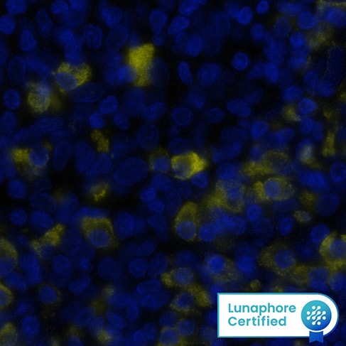
Detection of IgD in Human Tonsil via seqIF™ staining on COMET™
IgD was detected in immersion fixed paraffin-embedded sections of human Tonsil using Mouse Anti-Human IgD, Monoclonal Antibody (Catalog # MAB9857) at 0.5ug/mL at 37 ° Celsius for 2 minutes. Before incubation with the primary antibody, tissue underwent an all-in-one dewaxing and antigen retrieval preprocessing using PreTreatment Module (PT Module) and Dewax and HIER Buffer H (pH 9; Epredia Catalog # TA-999-DHBH). Tissue was stained using the Alexa Fluor™ 555 Goat anti-Mouse IgG Secondary Antibody at 1:100 at 37 ° Celsius for 2 minutes. (Yellow; Lunaphore Catalog # DR555MS) and counterstained with DAPI (blue; Lunaphore Catalog # DR100). Specific staining was localized to the cytoplasm. Protocol available in COMET™ Panel Builder.Detection of IgD in Human PBMCs by Flow Cytometry.
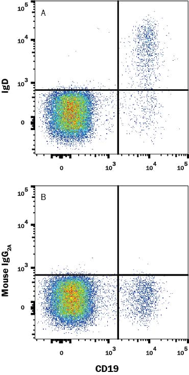
Human peripheral blood mononuclear cells (PBMCs) were stained with Mouse Anti-Human CD19 PE-conjugated Monoclonal Antibody (FAB4867P) and either (A) Mouse Anti-Human IgD Monoclonal Antibody (Catalog # MAB9857) or (B) Mouse IgG2A Isotype Control (MAB003) followed by Allophycocyanin-conjugated Anti-Mouse IgG Secondary Antibody (F0101B). View our protocol for Staining Membrane-associated Proteins.Detection of IgD in Human Tonsil.
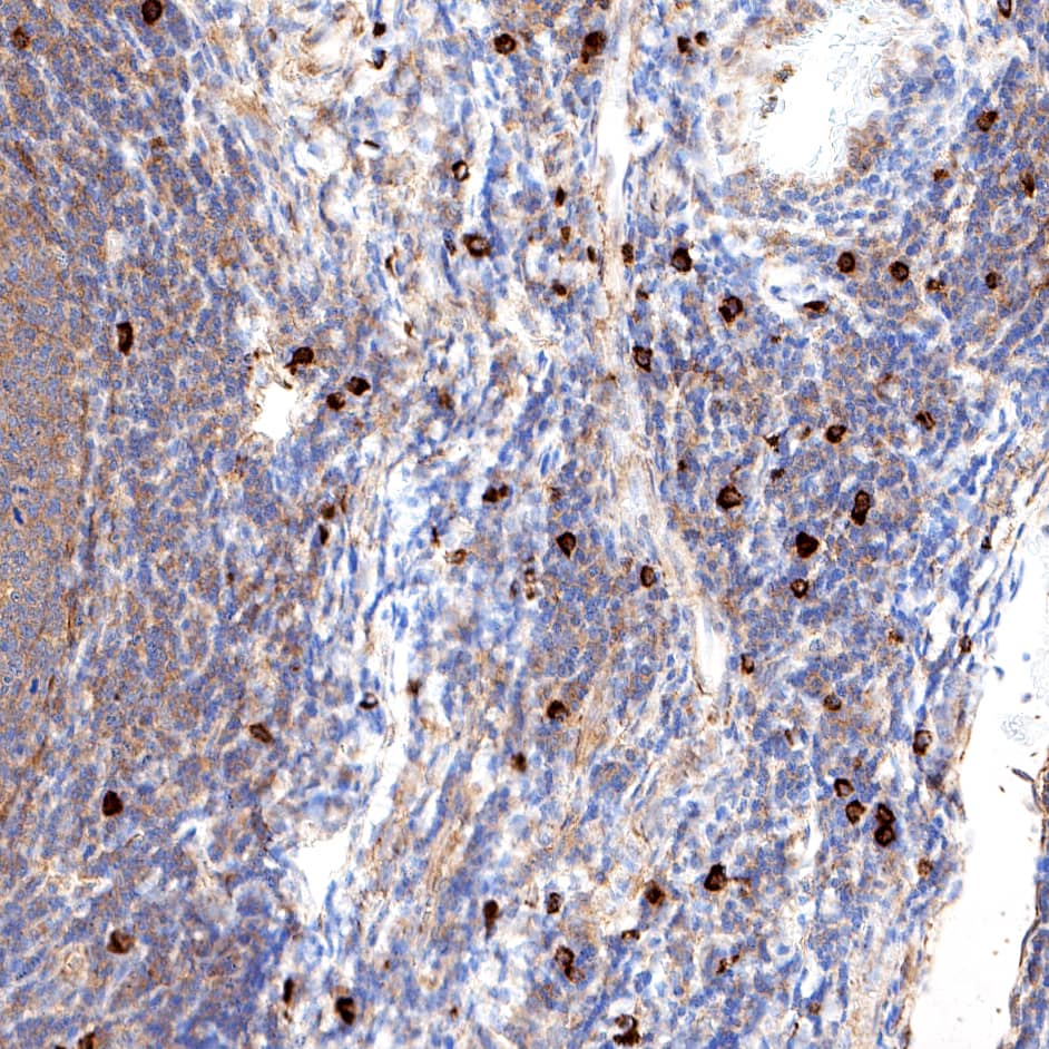
IgD was detected in immersion fixed paraffin-embedded sections of human tonsil using Mouse Anti-Human IgD Monoclonal Antibody (Catalog # MAB9857) at 5 µg/ml for 1 hour at room temperature followed by incubation with the HRP-conjugated Anti-Mouse IgG Secondary Antibody (Catalog # HAF007) or the Anti-Mouse IgG VisUCyte™ HRP Polymer Antibody (Catalog # VC001). Before incubation with the primary antibody, tissue was subjected to heat-induced epitope retrieval using VisUCyte Antigen Retrieval Reagent-Basic (Catalog # VCTS021). Tissue was stained using DAB (brown) and counterstained with hematoxylin (blue). Specific staining was localized to the membrane of B cells. View our protocol for Chromogenic IHC Staining of Paraffin-embedded Tissue Sections.Applications for Human IgD Antibody
Application
Recommended Usage
CyTOF-ready
Ready to be labeled using established conjugation methods. No BSA or other carrier proteins that could interfere with conjugation.
Flow Cytometry
0.25 µg/106 cells
Sample: Human peripheral blood mononuclear cells (PBMCs)
Sample: Human peripheral blood mononuclear cells (PBMCs)
Immunohistochemistry
3-25 µg/mL
Sample: Immersion fixed paraffin-embedded sections of human tonsil
Sample: Immersion fixed paraffin-embedded sections of human tonsil
Multiplex Immunofluorescence
0.5 µg/mL
Sample: Immersion fixed paraffin-embedded sections of human tonsil tissue
Sample: Immersion fixed paraffin-embedded sections of human tonsil tissue
Reviewed Applications
Read 1 review rated 5 using MAB9857 in the following applications:
Formulation, Preparation, and Storage
Purification
Protein A or G purified from hybridoma culture supernatant
Reconstitution
Reconstitute at 0.5 mg/mL in sterile PBS. For liquid material, refer to CoA for concentration.
Formulation
Lyophilized from a 0.2 μm filtered solution in PBS with Trehalose. See Certificate of Analysis for details.
*Small pack size (-SP) is supplied either lyophilized or as a 0.2 µm filtered solution in PBS.
*Small pack size (-SP) is supplied either lyophilized or as a 0.2 µm filtered solution in PBS.
Shipping
Lyophilized product is shipped at ambient temperature. Liquid small pack size (-SP) is shipped with polar packs. Upon receipt, store immediately at the temperature recommended below.
Stability & Storage
Use a manual defrost freezer and avoid repeated freeze-thaw cycles.
- 12 months from date of receipt, -20 to -70 °C as supplied.
- 1 month, 2 to 8 °C under sterile conditions after reconstitution.
- 6 months, -20 to -70 °C under sterile conditions after reconstitution.
Background: IgD
Long Name
Immunoglobulin D
Alternate Names
Igh-5, Ighd, Immunoglobulin Delta
Gene Symbol
IGHD
Additional IgD Products
Product Documents for Human IgD Antibody
Product Specific Notices for Human IgD Antibody
For research use only
Loading...
Loading...
Loading...
Loading...
Loading...

