Human IGF-II R/IGF2R Antibody
R&D Systems, part of Bio-Techne | Catalog # AF2447


Key Product Details
Species Reactivity
Validated:
Cited:
Applications
Validated:
Cited:
Label
Antibody Source
Product Specifications
Immunogen
Ser1510-Phe2108
Accession # P11717
Specificity
Clonality
Host
Isotype
Endotoxin Level
Scientific Data Images for Human IGF-II R/IGF2R Antibody
Detection of IGF-II R/IGF2R in Human Monocytes by Flow Cytometry.
Human whole blood monocytes were stained with Goat Anti-Human IGF-II R/IGF2R Antigen Affinity-purified Polyclonal Antibody (Catalog # AF2447, filled histogram) or control antibody (Catalog # AB-108-C, open histogram), followed by Phycoerythrin-conjugated Anti-Goat IgG Secondary Antibody (Catalog # F0107).IGF-II R/IGF2R in A172 Human Cell Line.
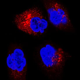
IGF-II R/IGF2R was detected in immersion fixed A172 human glioblastoma cell line using Goat Anti-Human IGF-II R/IGF2R Antigen Affinity-purified Polyclonal Antibody (Catalog # AF2447) at 5 µg/mL for 3 hours at room temperature. Cells were stained using the NorthernLights™ 557-conjugated Anti-Goat IgG Secondary Antibody (red; Catalog # NL001) and counterstained with DAPI (blue). Specific staining was localized to cytoplasm. View our protocol for Fluorescent ICC Staining of Cells on Coverslips.IGF-II R/IGF2R in A549 Human Cell Line.
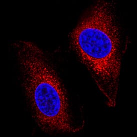
IGF-II R/IGF2R was detected in immersion fixed A549 human lung carcinoma cell line using Goat Anti-Human IGF-II R/IGF2R Antigen Affinity-purified Polyclonal Antibody (Catalog # AF2447) at 5 µg/mL for 3 hours at room temperature. Cells were stained using the NorthernLights™ 557-conjugated Anti-Goat IgG Secondary Antibody (red; Catalog # NL001) and counterstained with DAPI (blue). Specific staining was localized to cell surface and cytoplasm. View our protocol for Fluorescent ICC Staining of Cells on Coverslips.Applications for Human IGF-II R/IGF2R Antibody
Blockade of Receptor-ligand Interaction
CyTOF-ready
Flow Cytometry
Sample: Human whole blood monocytes
Immunocytochemistry
Sample: Fixed A172 human glioblastoma cell line and A549 human lung carcinoma cell line
Immunohistochemistry
Sample: Immersion fixed paraffin-embedded sections of human placenta
Western Blot
Sample: Recombinant Human IGF-II R/IGF2R (Catalog # 2447-GR)
Formulation, Preparation, and Storage
Purification
Reconstitution
Formulation
Shipping
Stability & Storage
- 12 months from date of receipt, -20 to -70 °C as supplied.
- 1 month, 2 to 8 °C under sterile conditions after reconstitution.
- 6 months, -20 to -70 °C under sterile conditions after reconstitution.
Background: IGF-II R/IGF2R
The type 2 insulin-like growth factor receptor (also known as cation-independent mannose-6 phosphate receptor/CI-MPR) is a 300 kDa member of the P-type lectin family of molecules. P-type lectins generate functional eukaryotic lysosomes by binding and sorting lysosomal enzymes expressing phosphorylated mannose residues (M6P) (1-3). IGF-II R is a type I transmembrane glycoprotein that contains a 2,264 amino acid (aa) extracellular region, a 23 aa transmembrane segment and a 124 aa cytoplasmic tail (4, 5). The extracellular region consists of 15 contiguous “binding” repeats of about 150 aa each. The odd-numbered repeats interact with “ligands” while the even-numbered repeats likely generate a nondisulfide homodimer in the membrane (1). Repeat #11 binds IGF-II, while repeats 3 and 9 bind mannose-6 phosphate; repeat #13 contains a fibronectin type II motif and assists in IGF-II binding (1, 2). In the extracellular region of IGF-II R expressed by R&D Systems (600 aa’s), human IGF-II R is 85% aa identical to both mouse and bovine IGF-II R. This expressed region includes binding repeats #11, 12, and 13. In addition to IGF-II, CI-MPR/IGF-II R binds non-M6P containing ligands such as retinoic acid, urokinase-type plasminogen-activator receptor and plasminogen, plus M6P‑containing molecules such as lysosomal enzymes, TGF-beta 1 precursor, proliferin, LIF, CD26, herpes simplex glycoprotein D, and granzymes A and B (2, 6). IGF-II R regulates many diverse biological functions that range from intracellular trafficking to the internalization of extracellular factors and modulation of cellular responses. It delivers newly synthesized M6P-tagged lysosomal enzymes from the trans-golgi network to endosomes, and facilitates the clearance of extracellular lysosomal and matrix degrading enzymes by internalization into clathrin-coated vesicles and delivery into endosomes. With respect to IGF-II biology, it would appear that IGF-II R is principally a regulator of local IGF-II levels, targeting IGF-II for destruction in lysosomes (2). However, some evidence suggests the receptor will signal via G‑proteins, an effect that has yet to be conclusively shown (6).
References
- Ghosh, P. et al. (2003) Nat. Rev. Mol. Cell. Biol. 4:202.
- Dahms, N.M. and M.K. Hancock (2002) Biochim. Biophys. Acta. 1572:317.
- Zaina, S. and J. Nilsson (2003) Curr. Opin. Lipidol. 14:483.
- Morgan, D.O. et al. (1987) Nature 329:301.
- Oshima, A. et al. (1988) J. Biol. Chem. 263:2553.
- Hawkes, C. and S. Kar (2004) Brain Res. Rev. 44:117.
Long Name
Alternate Names
Entrez Gene IDs
Gene Symbol
UniProt
Additional IGF-II R/IGF2R Products
Product Documents for Human IGF-II R/IGF2R Antibody
Product Specific Notices for Human IGF-II R/IGF2R Antibody
For research use only


