Human IRAK3 Antibody
R&D Systems, part of Bio-Techne | Catalog # AF6264

Key Product Details
Species Reactivity
Applications
Label
Antibody Source
Product Specifications
Immunogen
aa 1-596
Accession # Q9Y616
Specificity
Clonality
Host
Isotype
Scientific Data Images for Human IRAK3 Antibody
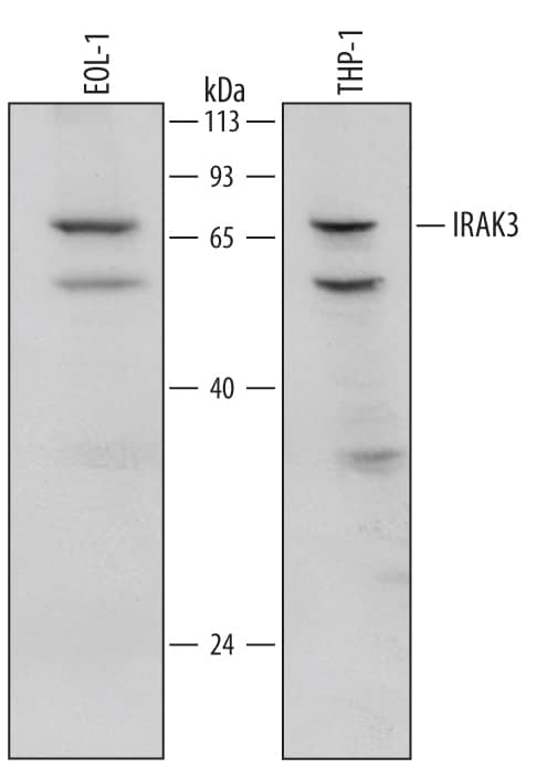
Detection of Human IRAK3 by Western Blot.
Western blot shows lysates of EOL-1 human acute myeloid (eosinophilic) leukemia cell line and THP-1 human acute monocytic leukemia cell line. PVDF Membrane was probed with 1 µg/mL of Sheep Anti-Human IRAK3 Antigen Affinity-purified Polyclonal Antibody (Catalog # AF6264) followed by HRP-conjugated Anti-Sheep IgG Secondary Antibody (HAF016). A specific band was detected for IRAK3 at approximately 68 kDa (as indicated). This experiment was conducted under reducing conditions and using Immunoblot Buffer Group 1.Detection of Human IRAK3 by Western Blot.
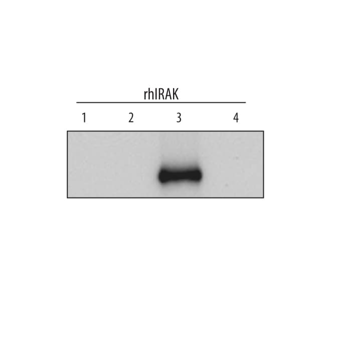
Western blot shows recombinant human IRAK1, recombinant human IRAK2, recombinant human IRAK3, and recombinant human IRAK4 (2 ng/lane). PVDF Membrane was probed with 1 µg/mL of Sheep Anti-Human IRAK3 Antigen Affinity-purified Polyclonal Antibody (Catalog # AF6264) followed by HRP-conjugated Anti-Sheep IgG Secondary Antibody (HAF016). This experiment was conducted under reducing conditions and using Immunoblot Buffer Group 1Detection of Human IRAK3 by Simple WesternTM.
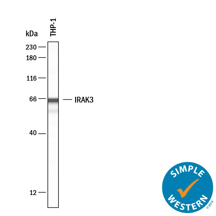
Simple Western lane view shows lysates of THP-1 human acute monocytic leukemia cell line, loaded at 0.2 mg/mL. A specific band was detected for IRAK3 at approximately 65 kDa (as indicated) using 20 µg/mL of Sheep Anti-Human IRAK3 Antigen Affinity-purified Polyclonal Antibody (Catalog # AF6264) . This experiment was conducted under reducing conditions and using the 12-230 kDa separation system.Applications for Human IRAK3 Antibody
Simple Western
Sample: Lysates of THP‑1 human acute monocytic leukemia cell line.
Western Blot
Sample: EOL‑1 human acute myeloid (eosinophilic) leukemia cell line and THP‑1 human acute monocytic leukemia cell line
Formulation, Preparation, and Storage
Purification
Reconstitution
Formulation
Shipping
Stability & Storage
- 12 months from date of receipt, -20 to -70 °C as supplied.
- 1 month, 2 to 8 °C under sterile conditions after reconstitution.
- 6 months, -20 to -70 °C under sterile conditions after reconstitution.
Background: IRAK3
IRAK3 (Interleukin-1 receptor-associated kinase 3; also IRAK-M) is a 68‑70 kDa member of the Pelle subfamily, TKL Ser/Thr protein kinase family of proteins. IRAK3 has limited expression, being mainly found in macrophages, eosinophils and respiratory epithelium, including type II alveolar cells. IRAK3 is a negative regulator of TLR signaling. Following TLR activation, it appears to form a complex with MyD88, IRAK1 and IRAK4 on the TLR cytoplasmic domain. This effectively blocks IRAK1/4 phosphorylation and inhibits NF kappaB and MAPK downstream activation. Human IRAK3 is 596 amino acids (aa) in length, contains one death domain (aa 41‑106) and a nonfunctional protein kinase domain (aa 171‑443). There are six Ser phosphorylation sites. There is one splice variant with a deletion of aa 45‑105.
Long Name
Alternate Names
Gene Symbol
UniProt
Additional IRAK3 Products
Product Documents for Human IRAK3 Antibody
Product Specific Notices for Human IRAK3 Antibody
For research use only


