Human IRF3 Antibody
R&D Systems, part of Bio-Techne | Catalog # AF4019

Key Product Details
Validated by
Species Reactivity
Applications
Label
Antibody Source
Product Specifications
Immunogen
Val206-Ser427
Accession # Q14653
Specificity
Clonality
Host
Isotype
Scientific Data Images for Human IRF3 Antibody
Detection of IRF3 in Daudi Human Cell Line by Flow Cytometry.
Daudi human Burkitt's lymphoma cell line was stained with Goat Anti-Human IRF3 Antigen Affinity-purified Polyclonal Antibody (Catalog # AF4019, filled histogram) or isotype control antibody (Catalog # AB-108-C, open histogram), followed by Phycoerythrin-conjugated Anti-Goat IgG Secondary Antibody (Catalog # F0107). To facilitate intracellular staining, cells were fixed with paraformaldehyde and permeabilized with saponin.Detection of Human IRF3 by Western Blot.
Western blot shows lysates of HeLa human cervical epithelial carcinoma cell line, U937 human histiocytic lymphoma cell line, Raji human Burkitt's lymphoma cell line, and Daudi human Burkitt's lymphoma cell line. PVDF membrane was probed with 1 µg/mL of Goat Anti-Human IRF3 Antigen Affinity-purified Polyclonal Antibody (Catalog # AF4019) followed by HRP-conjugated Anti-Goat IgG Secondary Antibody (Catalog # HAF019). A specific band was detected for IRF3 at approximately 50 kDa (as indicated). This experiment was conducted under reducing conditions and using Immunoblot Buffer Group 1.IRF3 in HeLa Human Cell Line.
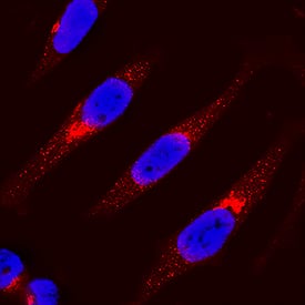
IRF3 was detected in immersion fixed HeLa human cervical epithelial carcinoma cell line using Goat Anti-Human IRF3 Antigen Affinity-purified Polyclonal Antibody (Catalog # AF4019) at 15 µg/mL for 3 hours at room temperature. Cells were stained using the NorthernLights™ 557-conjugated Anti-Goat IgG Secondary Antibody (red; Catalog # NL001) and counterstained with DAPI (blue). Specific staining was localized to cytoplasm. View our protocol for Fluorescent ICC Staining of Cells on Coverslips.Applications for Human IRF3 Antibody
CyTOF-ready
Immunocytochemistry
Sample: Immersion fixed HeLa human cervical epithelial carcinoma cell line
Intracellular Staining by Flow Cytometry
Sample: Daudi human Burkitt's lymphoma cell line fixed with paraformaldehyde and permeabilized with saponin
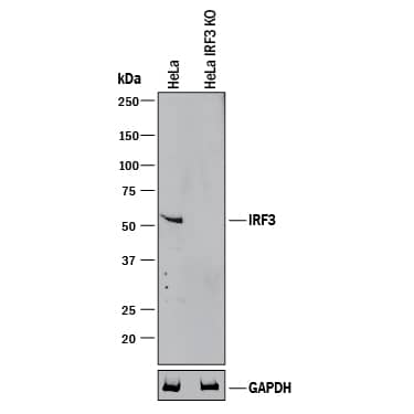
Knockout Validated
Simple Western
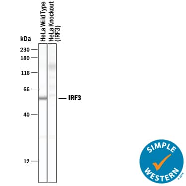
Sample: HeLa human cervical epithelial carcinoma cell line
Western Blot
Sample: HeLa human cervical epithelial carcinoma cell line, U937 human histiocytic lymphoma cell line, Raji human Burkitt's lymphoma cell line, and Daudi human Burkitt's lymphoma cell line
Formulation, Preparation, and Storage
Purification
Reconstitution
Formulation
Shipping
Stability & Storage
- 12 months from date of receipt, -20 to -70 °C as supplied.
- 1 month, 2 to 8 °C under sterile conditions after reconstitution.
- 6 months, -20 to -70 °C under sterile conditions after reconstitution.
Background: IRF3
Interferon response factor 3 (IRF3) is a 50 kDa, 427 amino acid, member of the IRF family of proteins. Human IRF3 is a transcription factor that binds the interferon-sensitive response element (ISRE) and is activated by Toll-like receptors, TLR3 and TLR4. Viral entry or engagement of TLR3 or TLR4 stimulates IRF3 phosphorylation, nuclear translocation and stimulation of IFN production. There are potential alternate splice forms affecting IRF3 structure, one including a deletion of aa 201‑327, a second with the same deletion and an alternate Met(147) start site, and a third with a 125 aa substitution of the C-terminal 100 aa. Over the region used as immunogen, aa 206‑427, human IRF3 is 76% and 83% identical to the corresponding mouse and porcine proteins, respectively.
Long Name
Alternate Names
Gene Symbol
UniProt
Additional IRF3 Products
Product Documents for Human IRF3 Antibody
Product Specific Notices for Human IRF3 Antibody
For research use only


