Human L1CAM Antibody
R&D Systems, part of Bio-Techne | Catalog # AF277


Key Product Details
Species Reactivity
Validated:
Human
Cited:
Human, Mouse
Applications
Validated:
Immunohistochemistry, Simple Western, Western Blot
Cited:
ELISA Development, Immunohistochemistry, Western Blot
Label
Unconjugated
Antibody Source
Polyclonal Goat IgG
Product Specifications
Immunogen
Mouse myeloma cell line NS0-derived recombinant human L1CAM
Ile20-Glu1120
Accession # CAA42508
Ile20-Glu1120
Accession # CAA42508
Specificity
Detects human L1CAM in direct ELISAs and Western blots. In direct ELISAs, approximately 10% cross‑reactivity with recombinant human (rh) ICAM-2 and rhICAM-3 is observed and less than 1% cross‑reactvity with rhCD31 and recombinant mouse VCAM-1 is observed.
Clonality
Polyclonal
Host
Goat
Isotype
IgG
Scientific Data Images for Human L1CAM Antibody
Detection of Human L1CAM by Western Blot.
Western blot shows lysates of HeLa human cervical epithelial carcinoma cell line and HDLM-2 human Hodgkin’s lymphoma cell line (negative control). PVDF membrane was probed with 0.25 µg/mL of Goat Anti-Human L1CAM Antigen Affinity-purified Polyclonal Antibody (Catalog # AF277) followed by HRP-conjugated Anti-Goat IgG Secondary Antibody (HAF017). A specific band was detected for L1CAM at approximately 250 kDa (as indicated). GAPDH (AF5718) is shown as a loading control. This experiment was conducted under reducing conditions and using Western Blot Buffer Group 1.Detection of Human and rat L1CAM by Western Blot.
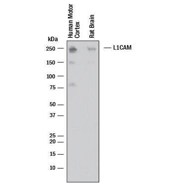
Western blot shows lysates of human brain (motor cortex) and rat brain. PVDF membrane was probed with 0.5 µg/mL of Goat Anti-Human L1CAM Antigen Affinity-purified Polyclonal Antibody (Catalog # AF277) followed by HRP-conjugated Anti-Goat IgG Secondary Antibody (HAF017). A specific band was detected for L1CAM at approximately 240 kDa (as indicated). This experiment was conducted under reducing conditions and using Western Blot Buffer Group 1.L1CAM in Human Brain.
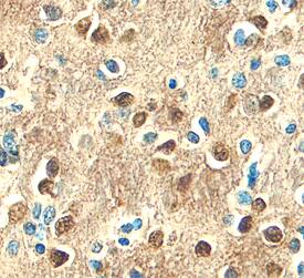
L1CAM was detected in immersion fixed paraffin-embedded sections of human brain (hippocampus) using Goat Anti-Human L1CAM Antigen Affinity-purified Poly-clonal Antibody (Catalog # AF277) at 10 µg/mL overnight at 4 °C. Before incubation with the primary antibody, tissue was subjected to heat-induced epitope retrieval using Antigen Retrieval Reagent-Basic (Catalog # CTS013). Tissue was stained using the Anti-Goat HRP-DAB Cell & Tissue Staining Kit (brown; Catalog # CTS008) and counter-stained with hematoxylin (blue). Specific staining was localized to plasma membranes and cytoplasm of neurons. View our protocol for Chromogenic IHC Staining of Paraffin-embedded Tissue Sections.Applications for Human L1CAM Antibody
Application
Recommended Usage
Immunohistochemistry
5-15 µg/mL
Sample: Immersion fixed paraffin-embedded sections of human brain (frontal cortex) and human brain (hippocampus) subjected to Antigen Retrieval Reagent-Basic (Catalog # CTS013)
Sample: Immersion fixed paraffin-embedded sections of human brain (frontal cortex) and human brain (hippocampus) subjected to Antigen Retrieval Reagent-Basic (Catalog # CTS013)
Simple Western
5 µg/mL
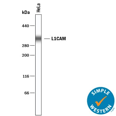
Sample: HeLa human cervical epithelial carcinoma cell line
Sample: HeLa human cervical epithelial carcinoma cell line
Western Blot
0.25 - 0.5 µg/mL
Sample: HeLa human cervical epithelial carcinoma cell line, human brain (motor cortex) and rat brain
Sample: HeLa human cervical epithelial carcinoma cell line, human brain (motor cortex) and rat brain
Reviewed Applications
Read 1 review rated 5 using AF277 in the following applications:
Formulation, Preparation, and Storage
Purification
Antigen Affinity-purified
Reconstitution
Reconstitute at 0.2 mg/mL in sterile PBS. For liquid material, refer to CoA for concentration.
Formulation
Lyophilized from a 0.2 μm filtered solution in PBS with Trehalose. *Small pack size (SP) is supplied either lyophilized or as a 0.2 µm filtered solution in PBS.
Shipping
Lyophilized product is shipped at ambient temperature. Liquid small pack size (-SP) is shipped with polar packs. Upon receipt, store immediately at the temperature recommended below.
Stability & Storage
Use a manual defrost freezer and avoid repeated freeze-thaw cycles.
- 12 months from date of receipt, -20 to -70 °C as supplied.
- 1 month, 2 to 8 °C under sterile conditions after reconstitution.
- 6 months, -20 to -70 °C under sterile conditions after reconstitution.
Background: L1CAM
Long Name
Cell Adhesion Molecule L1
Alternate Names
CAML1, CD171, HSAS, HSAS1, MASA, MIC5, NCAM-L1, S10, SPG1
Gene Symbol
L1CAM
UniProt
Additional L1CAM Products
Product Documents for Human L1CAM Antibody
Product Specific Notices for Human L1CAM Antibody
For research use only
Loading...
Loading...
Loading...
Loading...


