Human LAG-3 Antibody
R&D Systems, part of Bio-Techne | Catalog # MAB23194


Key Product Details
Validated by
Species Reactivity
Applications
Label
Antibody Source
Product Specifications
Immunogen
Met1-Leu450
Accession # P18627
Specificity
Clonality
Host
Isotype
Scientific Data Images for Human LAG-3 Antibody
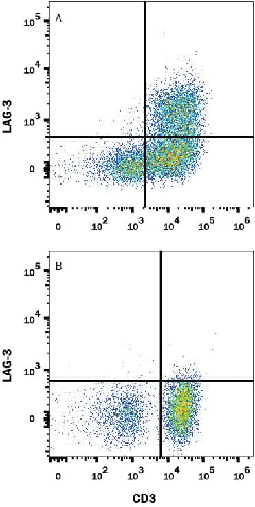
Detection of LAG‑3 in Human PBMCs by Flow Cytometry.
Human peripheral blood mononuclear cells (PBMCs) were either untreated (bottom panel) or treated with with 5 µg/mL PHA (top panel) for 5 days. PBMCs were stained with Mouse Anti-Human LAG-3 Monoclonal Antibody (Catalog # MAB23194) followed by Allophycocyanin-conjugated Anti-Mouse IgG Secondary Antibody (Catalog # F0101B) and Mouse Anti-Human CD3 epsilon PE-conjugated Monoclonal Antibody (Catalog # FAB100P). Quadrant markers were set based on isotype control antibody staining (Catalog # MAB003) (data not shown). View our protocol for Staining Membrane-associated Proteins.Detection of LAG-3 in HEK293 Human Cell Line Transfected with Human LAG-3 and eGFP by Flow Cytometry.
HEK293 human embryonic kidney cell line transfected with either (A) human LAG-3 or (B) irrelevant transfectants and eGFP was stained with Mouse Anti-Human LAG-3 Monoclonal Antibody (Catalog # MAB23194) followed by APC-conjugated Anti-Mouse IgG Secondary Antibody (Catalog # F0101B). Quadrant markers were set based on control antibody staining (Catalog # MAB003, data not shown). View our protocol for Staining Membrane-associated Proteins.Applications for Human LAG-3 Antibody
CyTOF-ready
Flow Cytometry
Sample: Human PBMC treated with PHA and HEK293 Human Cell Line Transfected with Human LAG-3 and eGFP
Formulation, Preparation, and Storage
Purification
Reconstitution
Formulation
Shipping
Stability & Storage
- 12 months from date of receipt, -20 to -70 °C as supplied.
- 1 month, 2 to 8 °C under sterile conditions after reconstitution.
- 6 months, -20 to -70 °C under sterile conditions after reconstitution.
Background: LAG-3
LAG-3 is an activation-induced molecule, expressed on activated T cells and NK cells, but not on resting T cells. Studies using LAG-3 -/- mice have shown significant delay of T cell apoptosis following antigen stimulation and increased size of memory T cells pool following infection (3, 4). It also has been reported that anti-LAG-3 antibodies up-regulate T cell activation by blocking interaction of LAG-3 and MHC class II. The study has demonstrated that LAG-3 is selectively expressed on activated CD4+CD25+ TReg cells and plays a role in their suppressive activity (5). This evidence indicated, unlike the interaction of CD4 with MHC class II that plays a positive role in T cell activation, LAG-3 binds to MHC class II and negatively regulates T cell activation through LAG-3 signaling. On the other hand, studies have shown that binding of LAG-3 to MHC class II molecules on antigen presenting cells induce maturation of dendritic cells and cytokine secretion by monocytes through MHC class II signal transduction (6). Taken together, LAG-3 may have two major functions, it negatively regulates T cells activation through LAG-3 signaling and stimulates antigen presenting cells which express MHC class II.
References
- Triebel, F. et al. (1990) J. Exp. Med. 171:1393.
- Baixeras, E. et al. (1992) J. Exp. Med 176:327.
- Workman, C.J. and D.A. Vignali (2003) Eur. J. Immunol. 33:970.
- Workman, C.J. et al. (2004) J. Immunol. 172:5450.
- Huang, C.T. et al. (2004) Immunity 21:503.
- Andreae, S. et al. (2003) Blood 102:2130.
Long Name
Alternate Names
Gene Symbol
UniProt
Additional LAG-3 Products
Product Documents for Human LAG-3 Antibody
Product Specific Notices for Human LAG-3 Antibody
For research use only
