Human LAG-3 Antibody
R&D Systems, part of Bio-Techne | Catalog # AF2319

Key Product Details
Validated by
Biological Validation
Species Reactivity
Validated:
Human
Cited:
Human
Applications
Validated:
CyTOF-reported, Flow Cytometry, Immunohistochemistry, Western Blot
Cited:
Flow Cytometry, Functional Assay, Immunocytochemistry, Western Blot
Label
Unconjugated
Antibody Source
Polyclonal Goat IgG
Product Specifications
Immunogen
Mouse myeloma cell line NS0-derived recombinant human LAG-3
Leu23-Leu450
Accession # P18627
Leu23-Leu450
Accession # P18627
Specificity
Detects human LAG-3 in direct ELISAs and Western blots. In direct ELISAs, less than 10% cross-reactivity with recombinant mouse LAG-3 is observed.
Clonality
Polyclonal
Host
Goat
Isotype
IgG
Scientific Data Images for Human LAG-3 Antibody
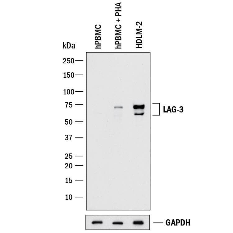
Detection of Human LAG‑3 by Western Blot.
Western blot shows lysates of human peripheral blood mononuclear cells (PBMC) untreated or treated (+) with 1 ug/mL PHA for 5 days and HDLM-2 human Hodgkin's lymphoma cell line. PVDF membrane was probed with 1 µg/mL of Goat Anti-Human LAG-3 Antigen Affinity-purified Polyclonal Antibody (Catalog # AF2319) followed by HRP-conjugated Anti-Goat IgG Secondary Antibody (Catalog # HAF017). Specific bands were detected for LAG-3 at approximately 60-75 kDa (as indicated). GAPDH (Catalog # MAB5718) is shown as a loading control. This experiment was conducted under reducing conditions and using Immunoblot Buffer Group 1.Detection of LAG‑3 in CD3+Human PBMCs by Flow Cytometry.
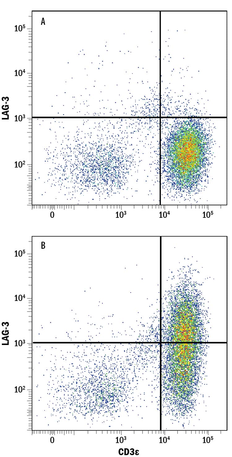
Human peripheral blood mononuclear cells (PBMCs) either (A) untreated or (B) treated with 1 ug/mL PHA for 5 days were stained with Goat Anti-Human LAG-3 Antigen Affinity-purified Polyclonal Antibody (Catalog # AF2319) followed by Allophycocyanin-conjugated Anti-Goat IgG Secondary Antibody (Catalog # F0108) and Mouse Anti-Human CD3e PE-conjugated Monoclonal Antibody (Catalog # FAB100P). Quadrant markers were set based on control antibody staining (Catalog # AB-108-C). View our protocol for Staining Membrane-associated Proteins.Detection of LAG-3 in HEK293 Human Cell Line Transfected with Human LAG-3 and eGFP by Flow Cytometry.
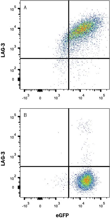
HEK293 human embryonic kidney cell line transfected with either (A) human LAG-3 or (B) irrelevant transfectants and eGFP was stained with Goat Anti-Human LAG-3 Antigen Affinity-purified Polyclonal Antibody (Catalog # AF2319) followed by Allophycocyanin-conjugated Anti-Goat IgG Secondary Antibody (Catalog # F0108). Quadrant markers were set based on control antibody staining(Catalog # AB-108-C, data not shown). View our protocol for Staining Membrane-associated Proteins.Applications for Human LAG-3 Antibody
Application
Recommended Usage
CyTOF-reported
Lowther, D.E. et al. (2016) JCI Insight 1:e85935. Ready to be labeled using established conjugation methods. No BSA or other carrier proteins that could interfere with conjugation.
Flow Cytometry
0.25 µg/106 cells
Sample: CD3+ human peripheral blood mononuclear cells (PBMCs) treated with PHA and HEK293 Human Cell Line Transfected with Human LAG-3 and eGFP
Sample: CD3+ human peripheral blood mononuclear cells (PBMCs) treated with PHA and HEK293 Human Cell Line Transfected with Human LAG-3 and eGFP
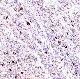
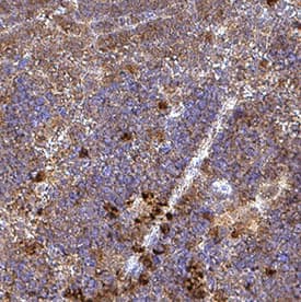
Immunohistochemistry
3-15 µg/mL
Sample: Immersion fixed paraffin-embedded sections of human spleen and mouse spleen
Sample: Immersion fixed paraffin-embedded sections of human spleen and mouse spleen
Western Blot
1 µg/mL
Sample: Human peripheral blood mononuclear cells (PBMC) treated with PHA and HDLM‑2 human Hodgkin's lymphoma cell line
Sample: Human peripheral blood mononuclear cells (PBMC) treated with PHA and HDLM‑2 human Hodgkin's lymphoma cell line
Formulation, Preparation, and Storage
Purification
Antigen Affinity-purified
Reconstitution
Reconstitute at 0.2 mg/mL in sterile PBS. For liquid material, refer to CoA for concentration.
Formulation
Lyophilized from a 0.2 μm filtered solution in PBS with Trehalose. *Small pack size (SP) is supplied either lyophilized or as a 0.2 µm filtered solution in PBS.
Shipping
Lyophilized product is shipped at ambient temperature. Liquid small pack size (-SP) is shipped with polar packs. Upon receipt, store immediately at the temperature recommended below.
Stability & Storage
Use a manual defrost freezer and avoid repeated freeze-thaw cycles.
- 12 months from date of receipt, -20 to -70 °C as supplied.
- 1 month, 2 to 8 °C under sterile conditions after reconstitution.
- 6 months, -20 to -70 °C under sterile conditions after reconstitution.
Background: LAG-3
References
- Triebel, F. et al. (1990) J. Exp. Med. 171:1393.
- Baixeras, E. et al. (1992) J. Exp. Med 176:327.
- Workman, C.J. and D.A. Vignali (2003) Eur. J. Immunol. 33:970.
- Workman, C.J. et al. (2004) J. Immunol. 172:5450.
- Huang, C.T. et al. (2004) Immunity 21:503.
- Andreae, S. et al. (2003) Blood 102:2130.
Long Name
Lymphocyte-activation Gene 3
Alternate Names
CD223, LAG3
Gene Symbol
LAG3
UniProt
Additional LAG-3 Products
Product Documents for Human LAG-3 Antibody
Product Specific Notices for Human LAG-3 Antibody
For research use only
Loading...
Loading...
Loading...
Loading...
Loading...
Loading...
