Human LIF Antibody
R&D Systems, part of Bio-Techne | Catalog # MAB250

Key Product Details
Validated by
Biological Validation
Species Reactivity
Validated:
Human
Cited:
Human
Applications
Validated:
Immunohistochemistry, Neutralization
Cited:
ELISA Development, Immunohistochemistry-Frozen, Neutralization, Western Blot
Label
Unconjugated
Antibody Source
Monoclonal Mouse IgG2B Clone # 9824
Product Specifications
Immunogen
E. coli-derived recombinant human LIF
Specificity
Detects human LIF in ELISAs and Western blots. In sandwich immunoassays, no significant cross-reactivity or interference with recombinant human (rh) IL-1 alpha, rhIL-1 beta, rhIL-2, rhIL-3, rhIL-4, rhIL-6, rhIL-7, rhIL-8, rhG-CSF, rhGM-CSF, rhOSM, rhTGF-beta 1, rhTNF-alpha, rhTNF-beta, recombinant mouse (rm) IL-1 beta, rmIL-3, rmIL-4, rmIL-5, rmIL-6, rmIL-7, rmGM-CSF, bovine (b) FGF acidic, bFGF basic, human (h) PDGF, porcine (p) PDGF, hTGF-beta 1, pTGF-beta 1.2, or pTGF-beta 2 is observed.
Clonality
Monoclonal
Host
Mouse
Isotype
IgG2B
Endotoxin Level
<0.10 EU per 1 μg of the antibody by the LAL method.
Scientific Data Images for Human LIF Antibody
Cell Proliferation Induced by LIF and Neutralization by Human LIF Antibody.
Recombinant Human LIF stimulates proliferation in the TF‑1 human erythroleukemic cell line in a dose-dependent manner (orange line). Proliferation elicited by 1.5 ng/mL Recombinant Human LIF is neutralized (green line) by increasing concentrations of Mouse Anti-Human LIF Monoclonal Antibody (Catalog # MAB250). The ND50 is typically 0.06-0.2 µg/mL.Detection of Human LIF by Western Blot
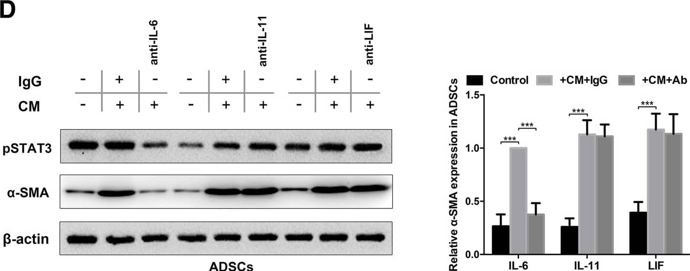
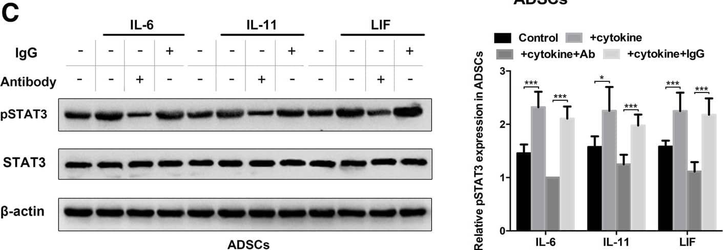
Autocrine IL-6 activates STAT3 signalling in lung cancer cell-induced epidural ADSCs. a Epidural ADSCs were pre-treated with CM from lung cancer cells for 48 h, and pSTAT3 and STAT3 expression levels were detected by western blotting. Epidural ADSCs cultured in untreated medium served as a control. b The effects of lung cancer cell CM on epidural ADSC proliferation were evaluated using the CCK-8 assay. Epidural ADSCs were treated with CM from one of four lung cancer cell lines, and the optical density of both groups at 450 nm was analysed. Data from three separate experiments are shown. c Western blot analysis of pSTAT3 and STAT3 in epidural ADSCs treated with either 10 ng/mL recombinant IL-6, 10 ng/mL recombinant IL-11 or 50 ng/mL recombinant LIF in the presence or absence of either neutralizing antibodies or isotype controls. Loading control, actin. d Western blot analysis of pSTAT3 and alpha-SMA expression in epidural ADSCs treated with lung cancer cell CM in the presence or absence of neutralizing antibodies against IL-6, IL-11 or LIF. Loading control, actin. *P < 0.05; **P < 0.01; ***P < 0.001 Image collected and cropped by CiteAb from the following publication (https://pubmed.ncbi.nlm.nih.gov/31196220), licensed under a CC-BY license. Not internally tested by R&D Systems.Detection of Human LIF by Western Blot
Autocrine IL-6 activates STAT3 signalling in lung cancer cell-induced epidural ADSCs. a Epidural ADSCs were pre-treated with CM from lung cancer cells for 48 h, and pSTAT3 and STAT3 expression levels were detected by western blotting. Epidural ADSCs cultured in untreated medium served as a control. b The effects of lung cancer cell CM on epidural ADSC proliferation were evaluated using the CCK-8 assay. Epidural ADSCs were treated with CM from one of four lung cancer cell lines, and the optical density of both groups at 450 nm was analysed. Data from three separate experiments are shown. c Western blot analysis of pSTAT3 and STAT3 in epidural ADSCs treated with either 10 ng/mL recombinant IL-6, 10 ng/mL recombinant IL-11 or 50 ng/mL recombinant LIF in the presence or absence of either neutralizing antibodies or isotype controls. Loading control, actin. d Western blot analysis of pSTAT3 and alpha-SMA expression in epidural ADSCs treated with lung cancer cell CM in the presence or absence of neutralizing antibodies against IL-6, IL-11 or LIF. Loading control, actin. *P < 0.05; **P < 0.01; ***P < 0.001 Image collected and cropped by CiteAb from the following publication (https://pubmed.ncbi.nlm.nih.gov/31196220), licensed under a CC-BY license. Not internally tested by R&D Systems.Applications for Human LIF Antibody
Application
Recommended Usage
Immunohistochemistry
8-25 µg/mL
Sample: Immersion fixed paraffin-embedded sections of human lung
Sample: Immersion fixed paraffin-embedded sections of human lung
Neutralization
Measured by its ability to neutralize LIF-induced proliferation in the TF‑1 human erythroleukemic cell line. Kitamura, T. et al. (1989) J. Cell Physiol. 140:323. The Neutralization Dose (ND50) is typically 0.06-0.2 µg/mL in the presence of 1.5 ng/mL Recombinant Human LIF. Human LIF Affinity-purified Polyclonal Antibody (Catalog # AF-250-NA) is recommended for neutralization.
Formulation, Preparation, and Storage
Purification
Protein A or G purified from ascites
Reconstitution
Reconstitute at 0.5 mg/mL in sterile PBS. For liquid material, refer to CoA for concentration.
Formulation
Lyophilized from a 0.2 μm filtered solution in PBS with Trehalose. *Small pack size (SP) is supplied either lyophilized or as a 0.2 µm filtered solution in PBS.
Shipping
Lyophilized product is shipped at ambient temperature. Liquid small pack size (-SP) is shipped with polar packs. Upon receipt, store immediately at the temperature recommended below.
Stability & Storage
Use a manual defrost freezer and avoid repeated freeze-thaw cycles.
- 12 months from date of receipt, -20 to -70 °C as supplied.
- 1 month, 2 to 8 °C under sterile conditions after reconstitution.
- 6 months, -20 to -70 °C under sterile conditions after reconstitution.
Background: LIF
References
- Van-Vlasselaev, P. et al. (1992) Prog. Growth Factor Res. 4:337.
- Gough, N.M. (1992) Growth Factors 7:175.
- Anegon, N.M, I. et al. (1991) J. Immunol.147:3973.
- Gillett, N.A. et al. (1993) Growth Factors 9:301.
- Banner, L. R. and P.H. Patterson (1994) Proc. Natl. Acad. Sci. 91:7109.
- Allen, E.H. et al. (1990) J. Cell. Physiol. 145:110.
- Lorenzo, J.A. et al. (1994) Clin.Immunol. Immunopathol. 70:260.
- Williams, R.L. et al. (1988) Nature 336:684.
- Baumann, H. et al. (1989) J. Immunol. 143:1163.
- Marshall, M.K. et al. (1994) Endocrinology 135:1412.
- Ludham, W.H. et al. (1994) Dev. Biol. 164:5283.
- Davis, S. et al. (1993) Science 260:18054.
- Gearing, D.P. et al. (1991) EMBO J. 10:28395.
- Gearing, D.P. et al. (1991) Science 255:1434.
- Hilton, D.J. and N.A. Nicola (1992) J. Biol. Chem. 267:102286.
- Hilton, D.J. et al. (1992) Ciba17. Foundation Symposium 167:2278.
Long Name
Leukemia Inhibitory Factor
Alternate Names
D Factor, Emfilermin, HILDA, MLPLI
Gene Symbol
LIF
Additional LIF Products
Product Documents for Human LIF Antibody
Product Specific Notices for Human LIF Antibody
For research use only
Loading...
Loading...
Loading...
Loading...


