Human MCM7 Antibody
R&D Systems, part of Bio-Techne | Catalog # MAB9217


Key Product Details
Species Reactivity
Applications
Label
Antibody Source
Product Specifications
Immunogen
Gly188-Ala328
Accession # P33993
Specificity
Clonality
Host
Isotype
Scientific Data Images for Human MCM7 Antibody
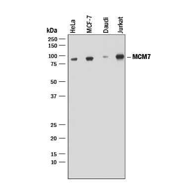
Detection of Human MCM7 by Western Blot.
Western blot shows lysates of HeLa human cervical epithelial carcinoma cell line, MCF-7 human breast cancer cell line, Daudi human Burkitt's lymphoma cell line, and Jurkat human acute T cell leukemia cell line. PVDF membrane was probed with 0.2 µg/mL of Rabbit Anti-Human MCM7 Monoclonal Antibody (Catalog # MAB9217) followed by HRP-conjugated Anti-Rabbit IgG Secondary Antibody (Catalog # HAF008). A specific band was detected for MCM7 at approximately 80 kDa (as indicated). This experiment was conducted under reducing conditions and using Immunoblot Buffer Group 1.MCM7 in HeLa Human Cell Line.
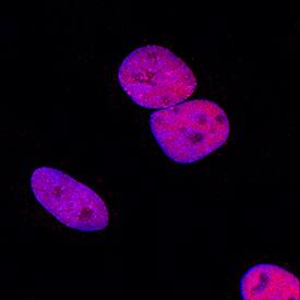
MCM7 was detected in immersion fixed HeLa human cervical epithelial carcinoma cell line using Rabbit Anti-Human MCM7 Monoclonal Antibody (Catalog # MAB9217) at 1 µg/mL for 3 hours at room temperature. Cells were stained using the NorthernLights™ 557-conjugated Anti-Rabbit IgG Secondary Antibody (red; Catalog # NL004) and counterstained with DAPI (blue). Specific staining was localized to nuclei. View our protocol for Fluorescent ICC Staining of Cells on Coverslips.MCM7 in Human Mesothelioma Tissue.
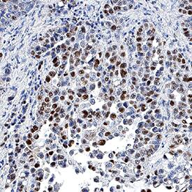
MCM7 was detected in immersion fixed paraffin-embedded sections of human mesothelioma tissue using Rabbit Anti-Human MCM7 Monoclonal Antibody (Catalog # MAB9217) at 3 µg/mL for 1 hour at room temperature followed by incubation with the Anti-Rabbit IgG VisUCyte™ HRP Polymer Antibody (Catalog # VC003). Tissue was stained using DAB (brown) and counterstained with hematoxylin (blue). Specific staining was localized to nuclei. View our protocol for IHC Staining with VisUCyte HRP Polymer Detection Reagents.Applications for Human MCM7 Antibody
Immunocytochemistry
Sample: Immersion fixed HeLa human cervical epithelial carcinoma cell line
Immunohistochemistry
Sample: Immersion fixed paraffin-embedded sections of human mesothelioma tissue
Intracellular Staining by Flow Cytometry
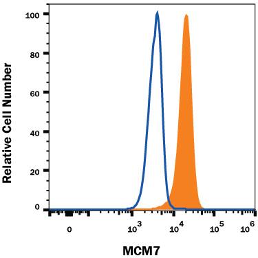
Sample: HeLa human cervical epithelial carcinoma cell line fixed and permeabilized with FlowX FoxP3 Fixation & Permeabilization Buffer Kit (Catalog # FC012)
Western Blot
Sample: HeLa human cervical epithelial carcinoma cell line, MCF‑7 human breast cancer cell line, Daudi human Burkitt's lymphoma cell line, and Jurkat human acute T cell leukemia cell line
Reviewed Applications
Read 1 review rated 5 using MAB9217 in the following applications:
Formulation, Preparation, and Storage
Purification
Reconstitution
Formulation
Shipping
Stability & Storage
- 12 months from date of receipt, -20 to -70 °C as supplied.
- 1 month, 2 to 8 °C under sterile conditions after reconstitution.
- 6 months, -20 to -70 °C under sterile conditions after reconstitution.
Background: MCM7
MCM7/minichromosome maintenance complex component 7, is a highly conserved mini-chromosome maintenance protein, with ATP binding, DNA helicase activity and single-stranded DNA binding. MCM7 forms a hexameric protein complex with MCM2, 4 and 6 proteins that has DNA helicase activity essential for the initiation of gene replication. MCM7 also binds to RAD17 to mediate ATR recruitment to damaged DNA. MCM7 expression is repressed in quiescent cells but induced in late G1 to S phase by growth factor stimulation. It is up-regulated in many cancers including colorectal , T cell lymphomas, glioblastoma, and has prognostic value for hepatocellular carcinoma and gastric adenocarcinoma.
Long Name
Alternate Names
Gene Symbol
UniProt
Additional MCM7 Products
Product Documents for Human MCM7 Antibody
Product Specific Notices for Human MCM7 Antibody
For research use only


