Human MINA Antibody
R&D Systems, part of Bio-Techne | Catalog # MAB7476


Key Product Details
Species Reactivity
Applications
Label
Antibody Source
Product Specifications
Immunogen
Met1-Gly192
Accession # Q8IUF8
Specificity
Clonality
Host
Isotype
Scientific Data Images for Human MINA Antibody
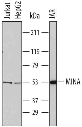
Detection of Human MINA by Western Blot.
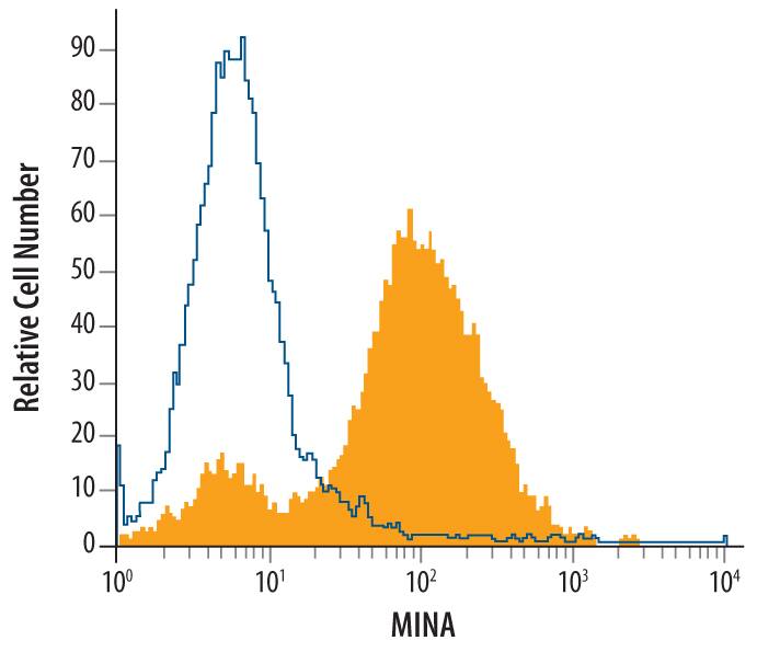
Western blot shows lysates of Jurkat human acute T cell leukemia cell line, HepG2 human hepatocellular carcinoma cell line, and JAR human choriocarcinoma cell line. PVDF membrane was probed with 2 µg/mL of Mouse Anti-Human MINA Monoclonal Antibody (Catalog # MAB7476) followed by HRP-conjugated Anti-Mouse IgG Secondary Antibody (Catalog # HAF007). A specific band was detected for MINA at approximately 53 kDa (as indicated). This experiment was conducted under reducing conditions and using Immunoblot Buffer Group 1.Detection of MINA in Jurkat Human Cell Line by Flow Cytometry.
Jurkat human acute T cell leukemia cell line was stained with Mouse Anti-Human MINA Monoclonal Antibody (Catalog # MAB7476, filled histogram) or isotype control antibody (Catalog # MAB002, open histogram), followed by Phycoerythrin-conjugated Anti-Mouse IgG Secondary Antibody (Catalog # F0102B). To facilitate intracellular staining, cells were fixed with paraformaldehyde and permeabilized with saponin.Applications for Human MINA Antibody
CyTOF-ready
Intracellular Staining by Flow Cytometry
Sample: Jurkat human acute T cell leukemia cell line fixed with paraformaldehyde and permeabilized with saponin
Western Blot
Sample: Jurkat human acute T cell leukemia cell line, HepG2 human hepatocellular carcinoma cell line, and JAR human choriocarcinoma cell line
Formulation, Preparation, and Storage
Purification
Reconstitution
Formulation
Shipping
Stability & Storage
- 12 months from date of receipt, -20 to -70 °C as supplied.
- 1 month, 2 to 8 °C under sterile conditions after reconstitution.
- 6 months, -20 to -70 °C under sterile conditions after reconstitution.
Background: MINA
MINA (myc-induced nuclear antigen; also Mina53) is a 52-54 kDa member of both the MINA53/NO66 and Jumonji C family of proteins. It expression is associated with proliferating cells, and it has been found in cytoplasm, nucleus and nucleoli. MINA appears to be induced by c-myc, and synthesized by spermatogonia, occasional squamous epithelium, naïve T cells and select cancer cells. When expressed, MINA is reported to regulate expression of genes such as HGF, EGF-R and IL-4. It may exert its regulatory activity through an intrinsic demethylase function. Mouse MINA is 465 amino acids (aa) in length. It possesses one cupin (or enzyme‑associated) region (aa 51-363) that contains a JmjC domain (aa 139-271). There are two potential isoform variants that contain either a 12 aa substitution for aa 145‑465, or a 15 aa substitution for aa 228-465. Over aa 2-192, mouse MINA shares 92% and 82% aa sequence identity with rat and human MINA, respectively.
Long Name
Alternate Names
Gene Symbol
UniProt
Additional MINA Products
Product Documents for Human MINA Antibody
Product Specific Notices for Human MINA Antibody
For research use only
