Human/Mouse Heparan Sulfate 3‑O‑Sulfotransferase 4/HS3ST4 Antibody
R&D Systems, part of Bio-Techne | Catalog # AF6085

Key Product Details
Species Reactivity
Human, Mouse
Applications
Immunohistochemistry, Western Blot
Label
Unconjugated
Antibody Source
Polyclonal Sheep IgG
Product Specifications
Immunogen
E. coli-derived recombinant human Heparan Sulfate 3-O-Sulfotransferase4/HS3ST4
Gly184-Lys456
Accession # Q9Y661
Gly184-Lys456
Accession # Q9Y661
Specificity
Detects human and mouse Heparan Sulfate 3-O-Sulfotransferase4/HS3ST4 in direct ELISAs and Western blots.
Clonality
Polyclonal
Host
Sheep
Isotype
IgG
Scientific Data Images
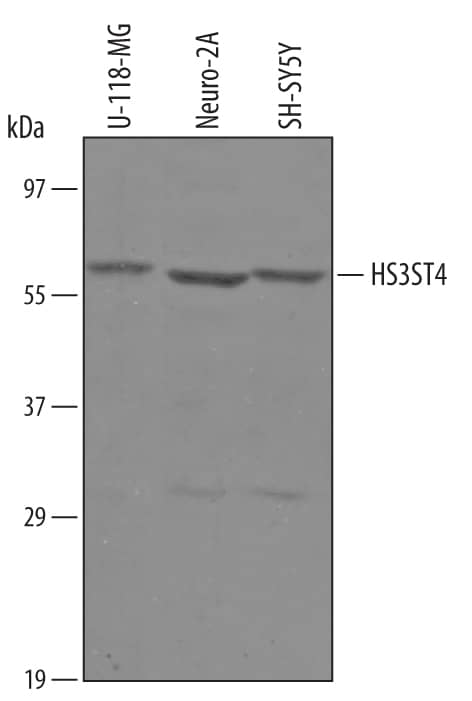
Detection of Human Sulfate 3-O-Sulfotransferase 4 /HS3ST4 by Western Blot.
Western blot shows lysates of U-118-MG human glioblastoma/astrocytoma cell line, Neuro-2A mouse neuroblastoma cell line, and SH-SY5Y human neuroblastoma cell line. PVDF Membrane was probed with 1 µg/mL of Sheep Anti-Human Sulfate 3-O-Sulfotransferase 4/HS3ST4 Antigen Affinity-purified Polyclonal Antibody (Catalog # AF6085) followed by HRP-conjugated Anti-Sheep IgG Secondary Antibody (Catalog # HAF016). A specific band was detected for Sulfate 3-O-Sulfotransferase 4/HS3ST4 at approximately 58 kDa (as indicated). This experiment was conducted under reducing conditions and using Immunoblot Buffer Group 8.Heparan Sulfate 3-O-Sulfotransferase 4/HS3ST4 in Human Brain.
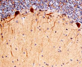
Heparan Sulfate 3-O-Sulfotransferase 4/HS3ST4 was detected in immersion fixed paraffin-embedded sections of human brain (cerebellum) using Sheep Anti-Human/Mouse Heparan Sulfate 3-O-Sulfotransferase 4/HS3ST4 Antigen Affinity-purified Polyclonal Antibody (Catalog # AF6085) at 3 µg/mL overnight at 4 °C. Before incubation with the primary antibody, tissue was subjected to heat-induced epitope retrieval using Antigen Retrieval Reagent-Basic (Catalog # CTS013). Tissue was stained using the Anti-Sheep HRP-DAB Cell & Tissue Staining Kit (brown; Catalog # CTS019) and counterstained with hematoxylin (blue). Specific staining was localized to Purkinje neurons. View our protocol for Chromogenic IHC Staining of Paraffin-embedded Tissue Sections.Applications
Application
Recommended Usage
Immunohistochemistry
5-15 µg/mL
Sample: Immersion fixed paraffin-embedded sections of human brain (cerebellum)
Sample: Immersion fixed paraffin-embedded sections of human brain (cerebellum)
Western Blot
1 µg/mL
Sample: U‑118‑MG human glioblastoma/astrocytoma cell line, Neuro‑2A mouse neuroblastoma cell line, and SH‑SY5Y human neuroblastoma cell line
Sample: U‑118‑MG human glioblastoma/astrocytoma cell line, Neuro‑2A mouse neuroblastoma cell line, and SH‑SY5Y human neuroblastoma cell line
Formulation, Preparation, and Storage
Purification
Antigen Affinity-purified
Reconstitution
Reconstitute at 0.2 mg/mL in sterile PBS. For liquid material, refer to CoA for concentration.
Formulation
Lyophilized from a 0.2 μm filtered solution in PBS with Trehalose. *Small pack size (SP) is supplied either lyophilized or as a 0.2 µm filtered solution in PBS.
Shipping
Lyophilized product is shipped at ambient temperature. Liquid small pack size (-SP) is shipped with polar packs. Upon receipt, store immediately at the temperature recommended below.
Stability & Storage
Use a manual defrost freezer and avoid repeated freeze-thaw cycles.
- 12 months from date of receipt, -20 to -70 °C as supplied.
- 1 month, 2 to 8 °C under sterile conditions after reconstitution.
- 6 months, -20 to -70 °C under sterile conditions after reconstitution.
Background: Heparan Sulfate 3-O-Sulfotransferase 4/HS3ST4
References
- Bernfield, M. et al. (1999) Annu. Rev. Biochem. 68:729.
- Esko, J.D. and Selleck, S.B. (2002) Annu. Rev. Biochem. 71:435.
- Shworak, N.W. et al. (1999) J. Biol. Chem. 274:5170.
- Xu, D. et al. (2005) Biochem. J. 386:451.
- Lawrence, R. et al. (2007) Matrix Biol. 26:442.
- Mochizuki, H. et al. (2003) J. Biol. Chem. 278:26780.
- Wu, Z.L. et al. (2004) J. Biol. Chem. 279:1861.
- Tiwari,V. et al. (2005) Biochem. Biophys. Res. Commun. 338:930.
- Wu, Z.L. et al. (2010) BMC Biotechnol. 10:11.
Alternate Names
3-OST-4
Entrez Gene IDs
9951 (Human)
Gene Symbol
HS3ST4
UniProt
Additional Heparan Sulfate 3-O-Sulfotransferase 4/HS3ST4 Products
Product Specific Notices
For research use only
Loading...
Loading...
Loading...
Loading...

