Human/Mouse MEF2C Antibody
R&D Systems, part of Bio-Techne | Catalog # AF6786

Key Product Details
Species Reactivity
Validated:
Cited:
Applications
Validated:
Cited:
Label
Antibody Source
Product Specifications
Immunogen
Ala135-Lys239
Accession # Q06413
Specificity
Clonality
Host
Isotype
Scientific Data Images for Human/Mouse MEF2C Antibody
Detection of Human MEF2C by Western Blot.
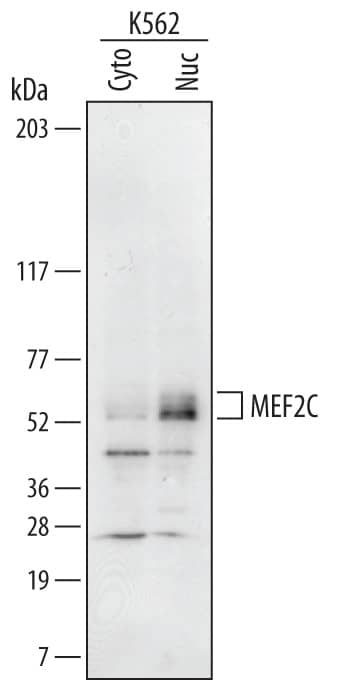
Western blot shows lysates of K562 human chronic myelogenous leukemia cell line. Gels were loaded with 20 µg of cytoplasmic (Cyto) and 10 µg of nuclear (Nuc) extracts. PVDF Membrane was probed with 0.2 µg/mL of Sheep Anti-Human/Mouse MEF2C Antigen Affinity-purified Polyclonal Antibody (Catalog # AF6786) followed by HRP-conjugated Anti-Sheep IgG Secondary Antibody (Catalog # HAF016). Specific bands were detected for MEF2C at approximately 52-60 kDa (as indicated). This experiment was conducted under reducing conditions and using Immunoblot Buffer Group 1.MEF2C in Human Brain.
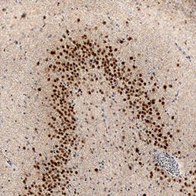
MEF2C was detected in immersion fixed paraffin-embedded sections of human brain (hippocampus) using Sheep Anti-Human/ Mouse MEF2C Antigen Affinity-purified Polyclonal Antibody (Catalog # AF6786) at 5 µg/mL overnight at 4 °C. Tissue was stained using the Anti-Sheep HRP-DAB Cell & Tissue Staining Kit (brown; Catalog # CTS019) and counterstained with hematoxylin (blue). Specific staining was localized to neuronal nuclei. View our protocol for Chromogenic IHC Staining of Paraffin-embedded Tissue Sections.MEF2C in Mouse Brain.
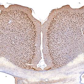
MEF2C was detected in perfusion fixed frozen sections of mouse brain (cortex) using Sheep Anti-Human/Mouse MEF2C Antigen Affinity-purified Polyclonal Antibody (Catalog # AF6786) at 1 µg/mL overnight at 4 °C. Tissue was stained using the Anti-Sheep HRP-DAB Cell & Tissue Staining Kit (brown; Catalog # CTS019) and counterstained with hematoxylin (blue). Specific staining was localized to neuronal nuclei. View our protocol for Chromogenic IHC Staining of Frozen Tissue Sections.Applications for Human/Mouse MEF2C Antibody
Immunohistochemistry
Sample: Immersion fixed paraffin-embedded sections of human brain (hippocampus) and perfusion fixed frozen sections of mouse brain (cortex)
Western Blot
Sample: K562 human chronic myelogenous leukemia cell line
Formulation, Preparation, and Storage
Purification
Reconstitution
Formulation
Shipping
Stability & Storage
- 12 months from date of receipt, -20 to -70 °C as supplied.
- 1 month, 2 to 8 °C under sterile conditions after reconstitution.
- 6 months, -20 to -70 °C under sterile conditions after reconstitution.
Background: MEF2C
MEF2C (Myocyte enhancer factor-2C) is a transcriptional activator that is a member of the MEF2 subfamily, MADS (MCM1, Agamous, Deficiens, Serum response factor) gene family of proteins. Although its predicted MW is 51 kDa, it runs anomalously at 57 kDa in SDS-PAGE. It is expressed in B cells, plus postmitotic neurons and skeletal muscle cells that are undergoing differentiation. Human MEF2C is 473 amino acids (aa) in length. It contains a MADS box for dimerization (aa 3-57), a DNA binding domain (aa 58-86), a beta-domain that enhances transcription (aa 271-278), a gamma-domain that, when phosphorylated, promotes transcriptional repression (aa 390-399), and seven acetylation sites plus multiple Ser/Thr phosphorylation sites. Sumoylation occurs on Lys391, and proteolytic cleavage of MEF2C occurs in neurons between Asp432Gly433. Multiple splice forms exist. Either individually or in combination, there may be a deletion of aa 271-278, 368-399 or 87-134, a 46 aa substitution for aa 107-134 and 87-134, and an alternative start site 35 aa upstream of the standard site. Over aa 135-239, human MEF2C shares 96% aa identity with mouse MEF2C.
Long Name
Alternate Names
Gene Symbol
UniProt
Additional MEF2C Products
Product Documents for Human/Mouse MEF2C Antibody
Product Specific Notices for Human/Mouse MEF2C Antibody
For research use only


