Human/Mouse Neogenin Antibody
R&D Systems, part of Bio-Techne | Catalog # AF1079

Key Product Details
Species Reactivity
Validated:
Cited:
Applications
Validated:
Cited:
Label
Antibody Source
Product Specifications
Immunogen
Ala42-Ile1033 (Asp442-Leu461 del)
Accession # NP_032710
Specificity
Clonality
Host
Isotype
Endotoxin Level
Scientific Data Images for Human/Mouse Neogenin Antibody
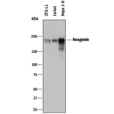
Detection of Human and Mouse Neogenin by Western Blot.
Western blot shows lysates of 3T3-L1 mouse embryonic fibroblast adipose-like cell line, Jurkat human acute T cell leukemia cell line, and Hepa 1-6 mouse hepatoma cell line. PVDF membrane was probed with 0.5 µg/mL of Goat Anti-Human/Mouse Neogenin Antigen Affinity-purified Polyclonal Antibody (Catalog # AF1079) followed by HRP-conjugated Anti-Goat IgG Secondary Antibody (Catalog # HAF017). A specific band was detected for Neogenin at approximately 230 kDa (as indicated). This experiment was conducted under reducing conditions and using Immunoblot Buffer Group 1.Neogenin in Mouse Embryo.
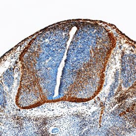
Neogenin was detected in perfusion fixed frozen sections of mouse embryo (15 d.p.c.) using Goat Anti-Human/Mouse Neogenin Antigen Affinity-purified Polyclonal Antibody (Catalog # AF1079) at 15 µg/mL overnight at 4 °C. Tissue was stained using the Anti-Goat HRP-DAB Cell & Tissue Staining Kit (brown; Catalog # CTS008) and counterstained with hematoxylin (blue). Specific staining was localized to developing neurons. View our protocol for Chromogenic IHC Staining of Frozen Tissue Sections.Detection of Mouse Neogenin by Simple WesternTM.
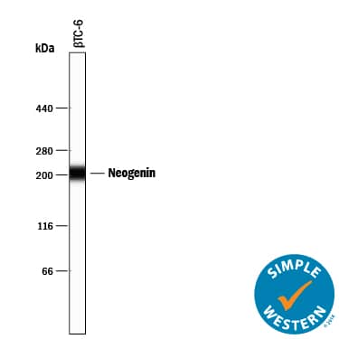
Simple Western lane view shows lysates of betaTC-6 mouse beta cell insulinoma cell line, loaded at 0.2 mg/mL. A specific band was detected for Neogenin at approximately 207 kDa (as indicated) using 10 µg/mL of Goat Anti-Human/Mouse Neogenin Antigen Affinity-purified Polyclonal Antibody (Catalog # AF1079) followed by 1:50 dilution of HRP-conjugated Anti-Goat IgG Secondary Antibody (Catalog # HAF109). This experiment was conducted under reducing conditions and using the 12-230 kDa separation system.Applications for Human/Mouse Neogenin Antibody
Blockade of Receptor-ligand Interaction
Immunohistochemistry
Sample: Perfusion fixed frozen sections of mouse embryo (15 d.p.c.)
Simple Western
Sample: betaTC‑6 mouse beta cell insulinoma cell line
Western Blot
Sample: 3T3‑L1 mouse embryonic fibroblast adipose-like cell line, Jurkat human acute T cell leukemia cell line, and Hepa 1‑6 mouse hepatoma cell line
Reviewed Applications
Read 1 review rated 4 using AF1079 in the following applications:
Formulation, Preparation, and Storage
Purification
Reconstitution
Formulation
Shipping
Stability & Storage
- 12 months from date of receipt, -20 to -70 °C as supplied.
- 1 month, 2 to 8 °C under sterile conditions after reconstitution.
- 6 months, -20 to -70 °C under sterile conditions after reconstitution.
Background: Neogenin
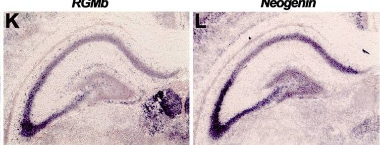
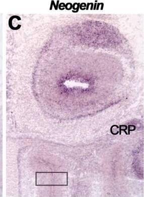
Neogenin (NEO) is a type I transmembrane protein that is crucial for axonal guidance and neuronal migration. It is also involved in regulating differentiation programs in many embryonic and adult tissues (1). Mouse NEO is widely expressed in adult tissues and is expressed throughout the mid to late stages of gestation, in both neuronal and non-neuronal tissues. It is a member of the immunoglobulin (Ig) superfamily and is closely related to deleted in colorectal cancer (DCC). Mouse NEO cDNA encodes a 1493 amino acid residue (aa) precursor with a putative 36 aa signal peptide, a 1100 aa extracellular domain with six Ig-like C2 type domains and three fibronectin type III domains, a 21 aa transmembrane domain, and a 345 aa cytoplasmic domain. At least five isoforms are produced in mice by alternative splicing. Mouse NEO shares 96%, 93%, and 86% aa sequence identity with rat, human, and chicken NEO, respectively. It also has 46% and 29% sequence homology with mouse DCC and C. elegans UNC40, a homolog of DCC. NEO and DCC, together with the UNC5 family of type I transmembrane proteins, are receptors for the netrin/UNC6 family of laminin-related bifunctional guidance molecules that both attract some axons and repel others (2, 3). In mouse, at least five netrins (netrin‑1,
‑3, -4, G1, and G2) have been identified (3-5). Mouse netrin-1 and netrin-3 have been shown to be ligands for mouse NEO.
References
- Keeling, S.L. et al. (1997) Oncogene 15:691.
- Hong, K. et al. (1999) Cell 97:927.
- Livesey, F.J. (1999) Cell Mol. Life Sci. 56:62.
- Nakashiba, T. et al. (2000) J. Neurosci. 20:6540.
- Nakashiba, T. et al. (2002) Mech. Dev. 111:47.
Alternate Names
Gene Symbol
UniProt
Additional Neogenin Products
Product Documents for Human/Mouse Neogenin Antibody
Product Specific Notices for Human/Mouse Neogenin Antibody
This product or the use of this product is covered by U.S. Patents owned by The Regents of the University of California. This product is for research use only and is not to be used for commercial purposes. Use of this product to produce products for sale or for diagnostic, therapeutic or drug discovery purposes is prohibited. In order to obtain a license to use this product for such purposes, contact The Regents of the University of California.
U.S. Patent # 5,939,271, 6,277,585, and other U.S. and international patents pending.
For research use only
