Human/Mouse PEA-15 Antibody
R&D Systems, part of Bio-Techne | Catalog # AF5588

Key Product Details
Validated by
Biological Validation
Species Reactivity
Human, Mouse
Applications
Immunohistochemistry, Western Blot
Label
Unconjugated
Antibody Source
Polyclonal Sheep IgG
Product Specifications
Immunogen
E. coli-derived recombinant human PEA-15
Ala2-Ala130
Accession # Q15121
Ala2-Ala130
Accession # Q15121
Specificity
Detects human and mouse PEA-15 in direct ELISAs and Western blots.
Clonality
Polyclonal
Host
Sheep
Isotype
IgG
Scientific Data Images for Human/Mouse PEA-15 Antibody
Detection of Human PEA-15 by Western Blot.
Western blot shows lysates of A172 human glioblastoma cell line and human cortex tissue. PVDF membrane was probed with 1 µg/mL of Sheep Anti-Human/Mouse PEA-15 Antigen Affinity-purified Polyclonal Antibody (Catalog # AF5588) followed by HRP-conjugated Anti-Sheep IgG Secondary Antibody (Catalog # HAF016). A specific band was detected for PEA-15 at approximately 15 kDa (as indicated). This experiment was conducted under reducing conditions and using Immunoblot Buffer Group 8.PEA‑15 in Human Brain.
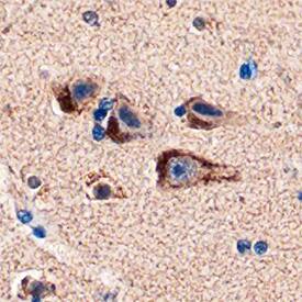
PEA-15 was detected in immersion fixed paraffin-embedded sections of human brain (cortex) using Sheep Anti-Human/Mouse PEA-15 Antigen Affinity-purified Polyclonal Antibody (Catalog # AF5588) at 1 µg/mL overnight at 4 °C. Tissue was stained using the Anti-Sheep HRP-DAB Cell & Tissue Staining Kit (brown; Catalog # CTS019) and counterstained with hematoxylin (blue). Specific staining was localized to neurons. View our protocol for Chromogenic IHC Staining of Paraffin-embedded Tissue Sections.Detection of Human PEA-15 by Western Blot
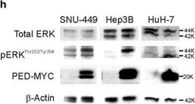
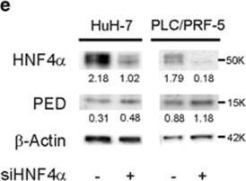
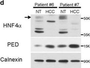
PED is inversely correlated to HNF4 alpha expression. (a) SNU-449 cells were co-transfected with 100 ng of pPED477 PED promoter-luciferase or pGL3 basic construct and treated with siRNA against HNF4 alpha or siRNA control. Luciferase activity was normalized for Renilla activity and is presented as mean±S.D. A representative experiment in triplicate is shown. (b,c) PED expression levels in HCC samples (b; n=59) or corresponding non-tumoral liver tissue (c, n=59) were correlated with HNF4 alpha expression. Correlation was calculated by Spearman test. Data are reported as probe intensity of an mRNA transcriptome array. (d) Western blot analysis for HNF4 alpha and PED in two HCC patient tumor samples and their corresponding non-tumoral (NT) tissues. Calnexin was used as loading control. Arrow: canonical full length HNF4 alpha (52 kDa); other bands are isoforms or truncated forms of the protein. (e,f) HuH-7 and PLC/PRF/5 cell lines were transfected with siRNA against HNF4 alpha (siHNF4 alpha) or siRNA control. After 72 h the protein expression of HNF4 alpha and PED was measured by western blot (e) and beta-actin served as control. mRNA expression was measured by qPCR (f) using RNA 18 S as internal control at 48 h for HuH-7 and 72 h for PLC/PRF/5. Data are reported as mean±S.D. of two independent experiments performed in triplicate. (g) SNU-449 cells were transfected with siRNA against HNF4 alpha or siRNA against PED alone or in combination, or siRNA control, as indicated. Migration was assessed by CIM plate with xCELLigence apparatus after 12 h and 24 h. Data are reported as mean±S.D. of two independent experiments performed in triplicate. (h) Western blot analysis of pERKThr202/Tyr204 and ERK in SNU-449, Hep3B and HuH-7 cell lines transfected with PED-MYC. beta-Actin was used as loading control. (i) pERKThr202/Tyr204 expression in two HCC patients and their non-tumoral counterpart. Calnexin was used as loading control. *P<0.05, **P<0.01, ****P<0.0001 Image collected and cropped by CiteAb from the following publication (https://www.nature.com/articles/cddis2017512), licensed under a CC-BY license. Not internally tested by R&D Systems.Applications for Human/Mouse PEA-15 Antibody
Application
Recommended Usage
Immunohistochemistry
5-15 µg/mL
Sample: Immersion fixed paraffin-embedded sections of human brain (cortex)
Sample: Immersion fixed paraffin-embedded sections of human brain (cortex)
Western Blot
1 µg/mL
Sample: A172 human glioblastoma cell line and human cortex tissue
Sample: A172 human glioblastoma cell line and human cortex tissue
Formulation, Preparation, and Storage
Purification
Antigen Affinity-purified
Reconstitution
Reconstitute at 0.2 mg/mL in sterile PBS. For liquid material, refer to CoA for concentration.
Formulation
Lyophilized from a 0.2 μm filtered solution in PBS with Trehalose. *Small pack size (SP) is supplied either lyophilized or as a 0.2 µm filtered solution in PBS.
Shipping
Lyophilized product is shipped at ambient temperature. Liquid small pack size (-SP) is shipped with polar packs. Upon receipt, store immediately at the temperature recommended below.
Stability & Storage
Use a manual defrost freezer and avoid repeated freeze-thaw cycles.
- 12 months from date of receipt, -20 to -70 °C as supplied.
- 1 month, 2 to 8 °C under sterile conditions after reconstitution.
- 6 months, -20 to -70 °C under sterile conditions after reconstitution.
Background: PEA-15
Long Name
Phosphoprotein Enriched in Astrocytes 15
Alternate Names
HMAT1, HUMMAT1H, MAT1H, PEA15, PED
Gene Symbol
PEA15
UniProt
Additional PEA-15 Products
Product Documents for Human/Mouse PEA-15 Antibody
Product Specific Notices for Human/Mouse PEA-15 Antibody
For research use only
Loading...
Loading...
Loading...
Loading...
