Human/Mouse Pin1 Antibody
R&D Systems, part of Bio-Techne | Catalog # MAB2294

Key Product Details
Species Reactivity
Validated:
Cited:
Applications
Validated:
Cited:
Label
Antibody Source
Product Specifications
Immunogen
Ala2-Glu163
Accession # Q13526
Specificity
Clonality
Host
Isotype
Scientific Data Images for Human/Mouse Pin1 Antibody
Detection of Human/Mouse Pin1 by Western Blot.
Western blot shows lysates of Balb/3T3 mouse embryonic fibroblast cell line, U2OS human osteosarcoma cell line, HeLa human cervical epithelial carcinoma cell line, MCF-7 human breast cancer cell line, and MDA-MB-453 human breast cancer cell line. PVDF membrane was probed with 0.5 µg/mL of Mouse Anti-Human/Mouse Pin1 Monoclonal Antibody (Catalog # MAB2294) followed by HRP-conjugated Anti-Mouse IgG Secondary Antibody (Catalog # HAF007). A specific band was detected for Pin1 at approximately 20 kDa (as indicated). This experiment was conducted under reducing conditions and using Immunoblot Buffer Group 1.Pin1 in MCF-7 Human Cell Line.
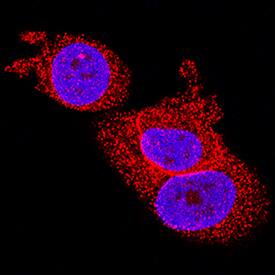
Pin1 was detected in immersion fixed MCF-7 human breast cancer cell line using Mouse Anti-Human/Mouse Pin1 Monoclonal Antibody (Catalog # MAB2294) at 8 µg/mL for 3 hours at room temperature. Cells were stained using the NorthernLights™ 557-conjugated Anti-Mouse IgG Secondary Antibody (red; Catalog # NL007) and counterstained with DAPI (blue). Specific staining was localized to nuclei and cytoplasm. View our protocol for Fluorescent ICC Staining of Cells on Coverslips.Pin1 in Human Breast Cancer Tissue.
Pin1 was detected in immersion fixed paraffin-embedded sections of human breast cancer tissue using Mouse Anti-Human/Mouse Pin1 Monoclonal Antibody (Catalog # MAB2294) at 25 µg/mL overnight at 4 °C. Tissue was stained using the Anti-Mouse HRP-DAB Cell & Tissue Staining Kit (brown; Catalog # CTS002) and counterstained with hematoxylin (blue). Specific labeling was localized to the cytoplasm of epithelial cells. View our protocol for Chromogenic IHC Staining of Paraffin-embedded Tissue Sections.Applications for Human/Mouse Pin1 Antibody
Immunocytochemistry
Sample: Immersion fixed MCF-7 human breast cancer cell line
Immunohistochemistry
Sample: Immersion fixed paraffin-embedded sections of human breast cancer tissue
Immunoprecipitation
Sample: MCF-7 human breast cancer cell line and HeLa human cervical epithelial carcinoma cell line, see our available Western blot detection antibodies
Western Blot
Sample: Balb/3T3 mouse embryonic fibroblast cell line, U2OS human osteosarcoma cell line, HeLa human cervical epithelial carcinoma cell line, MCF-7 human breast cancer cell line, and MDA-MB-453 human breast cancer cell line
Reviewed Applications
Read 1 review rated 5 using MAB2294 in the following applications:
Formulation, Preparation, and Storage
Purification
Reconstitution
Formulation
Shipping
Stability & Storage
- 12 months from date of receipt, -20 to -70 °C as supplied.
- 1 month, 2 to 8 °C under sterile conditions after reconstitution.
- 6 months, -20 to -70 °C under sterile conditions after reconstitution.
Background: Pin1
Pin1 is a peptidyl-prolyl isomerase (PPI) that targets phosphorylated Ser or Thr residues followed by a Pro (S/T-P). Isomerization of phosphorylated Ser or Thr residues may alter protein confirmation and, subsequently, modify activity. Pin1 is overexpressed in many human breast cancers, and has been shown to modify numerous proteins including p53,NF-kappa B, c-Jun, cyclin D1, and beta-catenin.
Long Name
Alternate Names
Entrez Gene IDs
Gene Symbol
UniProt
Additional Pin1 Products
Product Documents for Human/Mouse Pin1 Antibody
Product Specific Notices for Human/Mouse Pin1 Antibody
For research use only


