Human/Mouse/Rat CaM Kinase II Pan Specific Antibody
R&D Systems, part of Bio-Techne | Catalog # MAB7280

Key Product Details
Species Reactivity
Validated:
Cited:
Applications
Validated:
Cited:
Label
Antibody Source
Product Specifications
Immunogen
Ala448-Gln558
Accession # Q13555
Specificity
Clonality
Host
Isotype
Scientific Data Images for Human/Mouse/Rat CaM Kinase II Pan Specific Antibody
Detection of Human, Mouse, and Rat CaM Kinase II by Western Blot.
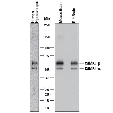
Western blot shows lysates of human brain (hippocampus) tissue, mouse brain tissue, and rat brain tissue. PVDF membrane was probed with 1 µg/mL of Mouse Anti-Human/Mouse/Rat CaM Kinase II Pan Specific Monoclonal Antibody (Catalog # MAB7280) followed by HRP-conjugated Anti-Mouse IgG Secondary Antibody (Catalog # HAF018). A specific band was detected for CaM Kinase II at approximately 50 and 60 kDa (as indicated). This experiment was conducted under reducing conditions and using Immunoblot Buffer Group 1.Detection of Recombinant Human CaM Kinase II alpha, beta gamma, and delta by Western Blot.
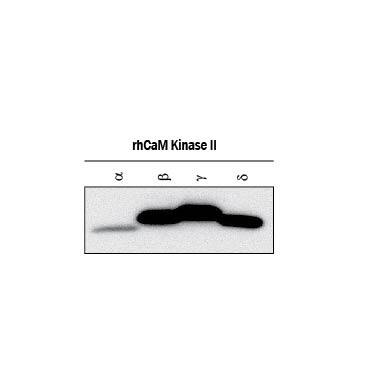
Western blot shows lysates of recombinant human CaM Kinase II a, recombinant human CaM Kinase II beta, recombinant human CaM Kinase II gamma, and recombinant human CaM Kinase II d. PVDF membrane was probed with 1 µg/mL of Mouse Anti-Human/Mouse/Rat CaM Kinase II Pan Specific Monoclonal Antibody (Catalog # MAB7280) followed by HRP-conjugated Anti-Sheep IgG Secondary Antibody (Catalog # HAF016). This experiment was conducted under reducing conditions and using Immunoblot Buffer Group 1.CaM Kinase II in PC‑3 Human Cell Line.
CaM Kinase II was detected in immersion fixed PC-3 human prostate cancer cell line using Mouse Anti-Human/Mouse/Rat CaM Kinase II Pan Specific Monoclonal Antibody (Catalog # MAB7280) at 8 µg/mL for 3 hours at room temperature. Cells were stained using the NorthernLights™ 557-conjugated Anti-Mouse IgG Secondary Antibody (red; Catalog # NL007) and counterstained with DAPI (blue). Specific staining was localized to cytoplasm. View our protocol for Fluorescent ICC Staining of Cells on Coverslips.Applications for Human/Mouse/Rat CaM Kinase II Pan Specific Antibody
Immunocytochemistry
Sample: Immersion fixed PC-3 human prostate cancer cell line and immersion fixed C2C12 mouse myoblast cell line
Immunohistochemistry
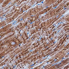
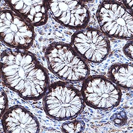
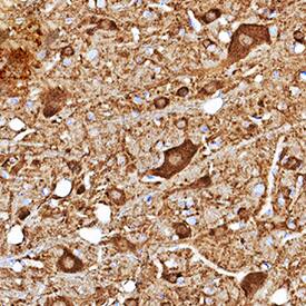
Sample: Immersion fixed paraffin-embedded sections of human heart, immersion fixed paraffin-embedded sections of human intestine, and perfusion fixed frozen sections of mouse brain (brainstem)
Western Blot
Sample: Human brain (hippocampus) tissue, Mouse brain tissue, Rat brain tissue, Recombinant human CaM Kinase II alpha, Recombinant human CaM Kinase II beta, Recombinant human CaM Kinase II gamma, and Recombinant human CaM Kinase II delta
Formulation, Preparation, and Storage
Purification
Reconstitution
Formulation
Shipping
Stability & Storage
- 12 months from date of receipt, -20 to -70 °C as supplied.
- 1 month, 2 to 8 °C under sterile conditions after reconstitution.
- 6 months, -20 to -70 °C under sterile conditions after reconstitution.
Background: CaM Kinase II
Calcium/calmodulin-dependent protein kinase type II gamma (CaMKII gamma) belongs to a family of multifunctional serine/threonine kinases activated in response to increases in intracellular calcium. There are 4 CaMKII isozymes, alpha, beta, gamma, and delta, and each can yield several isoforms through alternative splicing. CaMKII isoforms assemble into homo- or heteromultimeric holoenzymes composed of 8 to 12 subunits. The widely expressed CaMKII gamma from human, mouse, and rat share 100% aa sequence identity within aa 448-558 of isoform 1.
Long Name
Alternate Names
UniProt
Additional CaM Kinase II Products
Product Specific Notices for Human/Mouse/Rat CaM Kinase II Pan Specific Antibody
For research use only
