Human/Mouse/Rat Fascin Antibody
R&D Systems, part of Bio-Techne | Catalog # AF7745

Key Product Details
Validated by
Species Reactivity
Applications
Label
Antibody Source
Product Specifications
Immunogen
Met1-Tyr493
Accession # Q16658
Specificity
Clonality
Host
Isotype
Scientific Data Images for Human/Mouse/Rat Fascin Antibody
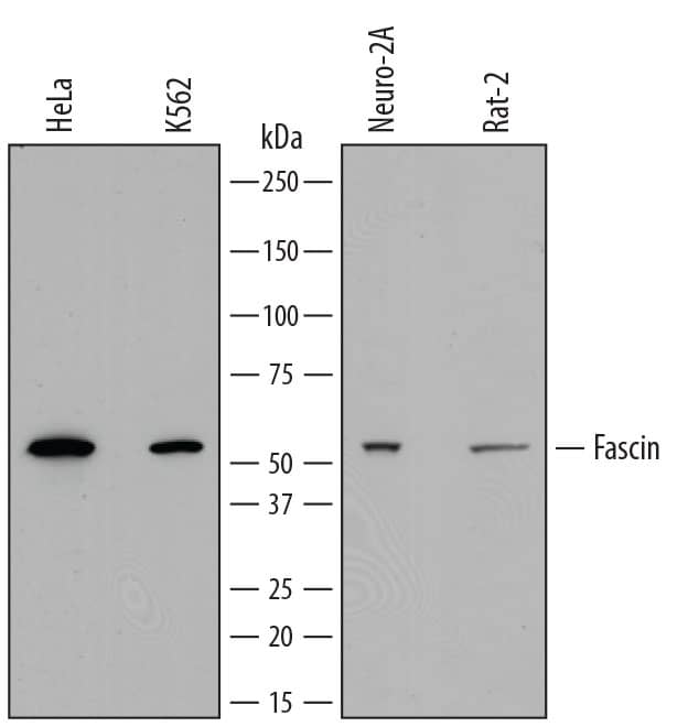
Detection of Human, Mouse, and Rat Fascin by Western Blot.
Western blot shows lysates of HeLa human cervical epithelial carcinoma cell line, K562 human chronic myelogenous leukemia cell line, Neuro-2A mouse neuroblastoma cell line, and Rat-2 rat thymidine kinase-deficient embryonic fibroblast cell line. PVDF membrane was probed with 0.4 µg/mL of Sheep Anti-Human/Mouse/Rat Fascin Antigen Affinity-purified Polyclonal Antibody (Catalog # AF7745) followed by HRP-conjugated Anti-Sheep IgG Secondary Antibody (Catalog # HAF016). A specific band was detected for Fascin at approximately 55 kDa (as indicated). This experiment was conducted under reducing conditions using Immunoblot Buffer Group 1./P>Western Blot Shows Human Fascin Specificity by Using Knockout Cell Line.
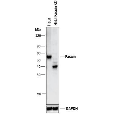
Western blot shows lysates of HeLa human cervical epithelial carcinoma parental cell line and Fascin knockout HeLa cell line (KO). PVDF membrane was probed with 0.4 µg/mL of Sheep Anti-Human/Mouse/Rat Fascin Antigen Affinity-purified Polyclonal Antibody (Catalog # AF7745) followed by HRP-conjugated Anti-Sheep IgG Secondary Antibody (Catalog # HAF016). A specific band was detected for Fascin at approximately 55 kDa (as indicated) in the parental HeLa cell line, but is not detectable in knockout HeLa cell line. GAPDH (Catalog # AF5718) is shown as a loading control. This experiment was conducted under reducing conditions and using Immunoblot Buffer Group 1. New adjunct appears with knockout cell line.Applications for Human/Mouse/Rat Fascin Antibody
Knockout Validated
Western Blot
Sample: HeLa human cervical epithelial carcinoma cell line, K562 human chronic myelogenous leukemia cell line, Neuro‑2A mouse neuroblastoma cell line, and Rat‑2 rat embryonic fibroblast cell line
Formulation, Preparation, and Storage
Purification
Reconstitution
Formulation
Shipping
Stability & Storage
- 12 months from date of receipt, -20 to -70 °C as supplied.
- 1 month, 2 to 8 °C under sterile conditions after reconstitution.
- 6 months, -20 to -70 °C under sterile conditions after reconstitution.
Background: Fascin
Fascin (that which creates fascicles [bundles] of actin; also known as 55 kDa actin-bundling protein, p55 and singed-like protein) is an intracellular 55-58 kDa member of the fascin family of proteins. It has a restricted expression pattern, being found in oligodendrocytes, select endothelium, cerebellar stellate neurons and blood, interdigitating, and thymic medullary dendritic cells. Fascin is found associated with actin in filopodia, and serves to coordinate and stabilize actin bundle formation, both in normal cells and tumor cells. In the latter cell type, filopodia have been renamed invadopodia, and their appearance is crucial for the creation of a stable platform that coordinates local matrix degradation. Human fascin is 493 amino acids (aa) in length. It contains an N-terminal fascin-like domain (aa 139-256) that contains part of one of two actin-binding sequences (aa 136-143), followed by two additional fascin-like domains (aa 260-378 and 383-493), the latter of which contains the second actin-binding sequence (aa 386-395). There are also two acetylation sites and two utilized phosphorylation sites at Ser38 and Ser39. Phosphorylation of the latter site inhibits fascin interaction with actin. At least two isoform variants may exist. One contains a 12 aa substitution for aa 427-493, while a second shows a deletion of aa 371-426. Full-length human fascin shares 97% aa sequence identity with mouse fascin. Two additional human fascins termed retinal and testis fascin have been identified. They are products of distinct genes and share 56% and 27% aa sequence identity with the standard (p55) fascin, respectively.
Alternate Names
Gene Symbol
UniProt
Additional Fascin Products
Product Documents for Human/Mouse/Rat Fascin Antibody
Product Specific Notices for Human/Mouse/Rat Fascin Antibody
For research use only

