Human/Mouse/Rat Histone Deacetylase 4/HDAC4 Antibody
R&D Systems, part of Bio-Techne | Catalog # AF6205

Key Product Details
Species Reactivity
Applications
Label
Antibody Source
Product Specifications
Immunogen
Met1-Gln68
Accession # P56524
Specificity
Clonality
Host
Isotype
Scientific Data Images for Human/Mouse/Rat Histone Deacetylase 4/HDAC4 Antibody
Detection of Human, Mouse, and Rat Histone Deacetylase 4/HDAC4 by Western Blot.
Western blot shows lysates of HeLa human cervical epithelial carcinoma cell line, Jurkat human acute T cell leukemia cell line, NIH-3T3 mouse embryonic fibroblast cell line, and NRK rat normal kidney cell line. PVDF Membrane was probed with 1 µg/mL of Human/Mouse/Rat Histone Deacetylase 4/HDAC4 Antigen Affinity-purified Polyclonal Antibody (Catalog # AF6205) followed by HRP-conjugated Anti-Sheep IgG Secondary Antibody (Catalog # HAF016). A specific band was detected for Histone Deacetylase 4/HDAC4 at approximately 140 kDa (as indicated). This experiment was conducted under reducing conditions and using Immunoblot Buffer Group 1.Histone Deacetylase 4/HDAC4 in MCF‑7 Human Cell Line.
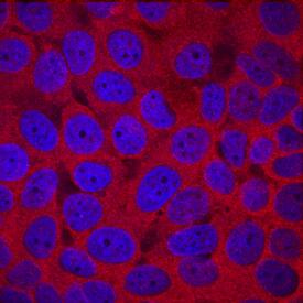
Histone Deacetylase 4/HDAC4 was detected in immersion fixed MCF-7 human breast cancer cell line using Sheep Anti-Human/Mouse/Rat Histone Deacetylase 4/ HDAC4 Antigen Affinity-purified Polyclonal Antibody (Catalog # AF6205) at 10 µg/mL for 3 hours at room temperature. Cells were stained using the NorthernLights™ 557-conjugated Anti-Sheep IgG Secondary Antibody (red; Catalog # NL010) and counterstained with DAPI (blue). Specific staining was localized to cytoplasm. View our protocol for Fluorescent ICC Staining of Cells on Coverslips.Applications for Human/Mouse/Rat Histone Deacetylase 4/HDAC4 Antibody
Immunocytochemistry
Sample: Immersion fixed MCF‑7 human breast cancer cell line
Western Blot
Sample: HeLa human cervical epithelial carcinoma cell line, Jurkat human acute T cell leukemia cell line, NIH‑3T3 mouse embryonic fibroblast cell line, and NRK rat normal kidney cell line
Formulation, Preparation, and Storage
Purification
Reconstitution
Formulation
Shipping
Stability & Storage
- 12 months from date of receipt, -20 to -70 °C as supplied.
- 1 month, 2 to 8 °C under sterile conditions after reconstitution.
- 6 months, -20 to -70 °C under sterile conditions after reconstitution.
Background: Histone Deacetylase 4/HDAC4
Histone Deacetylase 4 (HDAC4; also HD4) is a founding member of the class IIa subfamily, histone deacetylase family of transcriptional regulators. Although its predicted MW is 119 kDa, it runs anomalously at 140‑150 kDa in SDS‑PAGE which may be due to extensive phosphorylation plus SUMOylation. It has an N-terminal region that interacts with transcription factors and corepressors, and a C-terminal domain that shows deacetylase activity, thus repressing gene transcription. HDAC4 is found in osteoblasts, cardiac and skeletal muscle cells, and neurons. Human HDAC4 is 1084 amino acids (aa) in length. It contains one coiled-coil region (aa 67‑177), a SUMOylation site at Lys559, a histone deacetylation domain (aa 665‑993) and one NES (aa 1051‑1084). Caspase cleavage after Asp289 generates bioactive 97 and 34 kDa fragments. There is one potential splice variant that shows an alternative start site at Met118 coupled to a five aa insertion after Thr431. Over aa 1‑68, human HDAC4 shares 96% aa identity with mouse HDAC4.
Long Name
Alternate Names
Gene Symbol
UniProt
Additional Histone Deacetylase 4/HDAC4 Products
Product Documents for Human/Mouse/Rat Histone Deacetylase 4/HDAC4 Antibody
Product Specific Notices for Human/Mouse/Rat Histone Deacetylase 4/HDAC4 Antibody
For research use only

