Human/Mouse/Rat MBP Antibody
R&D Systems, part of Bio-Techne | Catalog # MAB42282

Key Product Details
Species Reactivity
Validated:
Cited:
Applications
Validated:
Cited:
Label
Antibody Source
Product Specifications
Immunogen
Accession # P02686
Specificity
Clonality
Host
Isotype
Scientific Data Images for Human/Mouse/Rat MBP Antibody
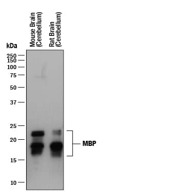
Detection of Human MBP by Western Blot.
Western blot shows lysates of mouse brain (cerebellum) tissue and rat brain (cerebellum). PVDF membrane was probed with 0.1 µg/mL of Mouse Anti-Human/Mouse/Rat MBP Monoclonal Antibody (Catalog # MAB42282) followed by HRP-conjugated Anti-Mouse IgG Secondary Antibody (Catalog # HAF018). Specific bands were detected for MBP at approximately 15-22 kDa (as indicated). This experiment was conducted under reducing conditions and using Immunoblot Buffer Group 1.Detection of Mouse and Rat MBP by Western Blot.
Western blot shows lysates of mouse brain (cerebellum) tissue and rat brain (cerebellum) tissue. PVDF membrane was probed with 0.1 µg/mL of Mouse Anti-Human/Mouse/Rat MBP Monoclonal Antibody (Catalog # MAB42282) followed by HRP-conjugated Anti-Mouse IgG Secondary Antibody (Catalog # HAF018). Specific bands were detected for MBP at approximately 15-22 kDa (as indicated). This experiment was conducted under reducing conditions and using Immunoblot Buffer Group 1.MBP in Rat Cortical Stem Cells.
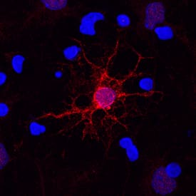
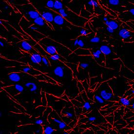
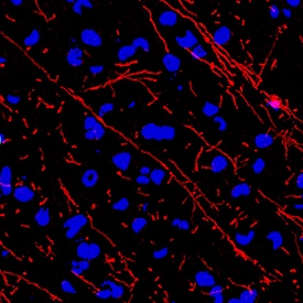
MBP was detected in immersion fixed rat cortical stem cells differentiated for 7 days to oligodendrocytes using Mouse Anti-Human/Mouse/Rat MBP Monoclonal Antibody (Catalog # MAB42282) at 10 µg/mL for 3 hours at room temperature. Cells were stained using the NorthernLights™ 557-conjugated Anti-Mouse IgG Secondary Antibody (red; Catalog # NL007) and counterstained with DAPI (blue). Specific staining was localized to cell surfaces, cytoplasm, and nuclei. View our protocol for Fluorescent ICC Staining of Stem Cells on Coverslips.Applications for Human/Mouse/Rat MBP Antibody
Immunocytochemistry
Sample: Immersion fixed rat cortical stem cells differentiated for 7 days to oligodendrocytes
Immunohistochemistry
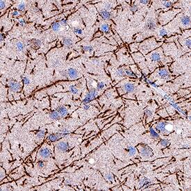
Sample: Perfusion fixed frozen sections of human brain, mouse brain, and rat brain
Western Blot
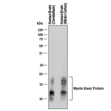
Sample: Human brain (cerebellum) tissue, Human brain (motor cortex) tissue, Mouse brain (cerebellum) tissue, and Rat brain (cerebellum) tissue
Reviewed Applications
Read 5 reviews rated 5 using MAB42282 in the following applications:
Formulation, Preparation, and Storage
Purification
Reconstitution
Formulation
Shipping
Stability & Storage
- 12 months from date of receipt, -20 to -70 °C as supplied.
- 1 month, 2 to 8 °C under sterile conditions after reconstitution.
- 6 months, -20 to -70 °C under sterile conditions after reconstitution.
Background: MBP
Myelin Basic Protein (MBP) is the most abundant protein component of the myelin membrane in the central nervous system. MBP has a role in both the formation and stabilization of this compact multilayer arrangement of bilayers. In vitro, MBP is suitable as a substrate for numerous protein kinases, including the ERK and p38 MAP kinases that phosphorylate MBP at T98.
Long Name
Alternate Names
Gene Symbol
UniProt
Additional MBP Products
Product Documents for Human/Mouse/Rat MBP Antibody
Product Specific Notices for Human/Mouse/Rat MBP Antibody
For research use only
