Human/Mouse Serpin F1/PEDF Antibody
R&D Systems, part of Bio-Techne | Catalog # AF1177


Key Product Details
Species Reactivity
Validated:
Cited:
Applications
Validated:
Cited:
Label
Antibody Source
Product Specifications
Immunogen
Asp44-Pro418
Accession # P36955
Specificity
Clonality
Host
Isotype
Scientific Data Images for Human/Mouse Serpin F1/PEDF Antibody
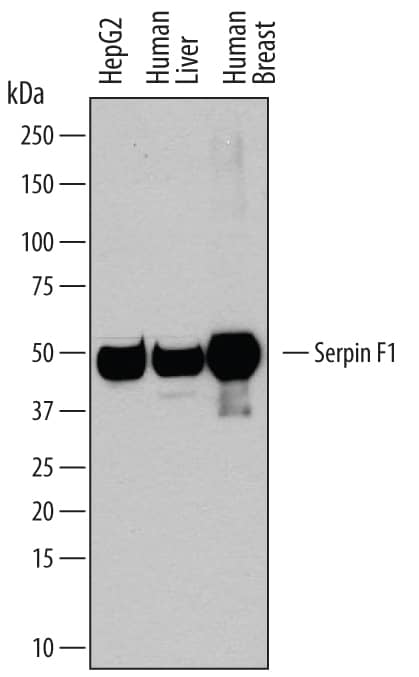
Detection of Human Serpin F1/PEDF by Western Blot.
Western blot shows lysates of HepG2 human hepatocellular carcinoma cell line, human liver tissue, and human breast tissue. PVDF membrane was probed with 0.2 µg/mL of Goat Anti-Human/Mouse Serpin F1/PEDF Antigen Affinity-purified Polyclonal Antibody (Catalog # AF1177) followed by HRP-conjugated Anti-Goat IgG Secondary Antibody (Catalog # HAF109). A specific band was detected for Serpin F1/PEDF at approximately 50 kDa (as indicated). This experiment was conducted under reducing conditions and using Immunoblot Buffer Group 1.Detection of Mouse Serpin F1/PEDF by Western Blot.
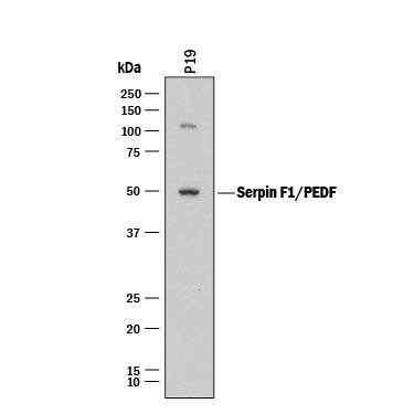
Western blot shows lysates of P19 mouse embryonal carcinoma cell line. PVDF membrane was probed with 2 µg/mL of Goat Anti-Human/Mouse Serpin F1/PEDF Antigen Affinity-purified Polyclonal Antibody (Catalog # AF1177) followed by HRP-conjugated Anti-Goat IgG Secondary Antibody (Catalog # HAF017). A specific band was detected for Serpin F1/PEDF at approximately 50 kDa (as indicated). This experiment was conducted under reducing conditions and using Immunoblot Buffer Group 1.Serpin F1/PEDF in Human Kidney.
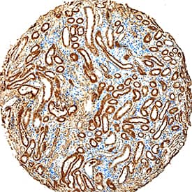
Serpin F1/PEDF was detected in immersion fixed paraffin-embedded sections of human kidney using Goat Anti-Human/Mouse Serpin F1/PEDF Antigen Affinity-purified Polyclonal Antibody (Catalog # AF1177) at 15 µg/mL overnight at 4 °C. Tissue was stained using the Anti-Goat HRP-DAB Cell & Tissue Staining Kit (brown; Catalog # CTS008) and counterstained with hematoxylin (blue). Specific staining was localized to convoluted tubules. View our protocol for Chromogenic IHC Staining of Paraffin-embedded Tissue Sections.Applications for Human/Mouse Serpin F1/PEDF Antibody
Immunohistochemistry
Sample: Immersion fixed paraffin-embedded sections of human skin and human kidney
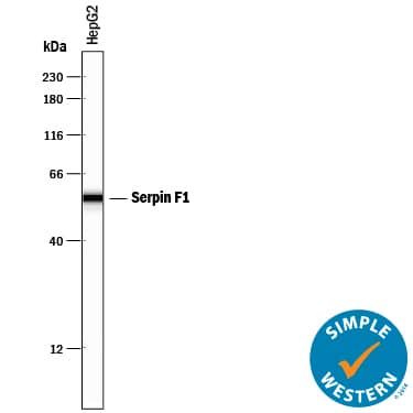
Simple Western
Sample: HepG2 human hepatocellular carcinoma cell line
Western Blot
Sample: HepG2 human hepatocellular carcinoma cell line, human liver tissue, human breast tissue, and P19 mouse embryonal carcinoma cell line
Formulation, Preparation, and Storage
Purification
Reconstitution
Formulation
Shipping
Stability & Storage
- 12 months from date of receipt, -20 to -70 °C as supplied.
- 1 month, 2 to 8 °C under sterile conditions after reconstitution.
- 6 months, -20 to -70 °C under sterile conditions after reconstitution.
Background: Serpin F1/PEDF
Serpin F1 (serine proteinase inhibitor-clade F #1; also PEDF, PIG35 and EPC-1) is a secreted, 50 kDa glycoprotein member of the clade F-subfamily, serpin superfamily of protease inhibitors. It is expressed by diverse cell types such as retinal pigment and breast epithelium, fibroblasts, astrocytes and hepatocytes. Serpin F1 circulates in blood and binds to type I collagen plus heparan sulfate. Although it is a serpin, it belongs to a class of serpins that are nonprotease inhibiting, and instead is known to possess potent antiangiogenic and neurotrophic activity. Human Serpin F1 is 418 amino acids (aa) in length. It contains a 19 aa signal sequence plus a 399 aa mature region that shows an NLS (aa 146-149), a neuroprotective motif (aa 354-359) and an antiangiogenesis segment (aa 387‑411). Phosphorylation occurs at multiple sites that affect bioactivity. Over aa 44‑418, human Serpin F1 shares 87% aa identity with mouse Serpin F1.
Long Name
Alternate Names
Gene Symbol
UniProt
Additional Serpin F1/PEDF Products
Product Documents for Human/Mouse Serpin F1/PEDF Antibody
Product Specific Notices for Human/Mouse Serpin F1/PEDF Antibody
For research use only


