Human N-Cadherin Propeptide Antibody
R&D Systems, part of Bio-Techne | Catalog # AF1388


Key Product Details
Species Reactivity
Validated:
Cited:
Applications
Validated:
Cited:
Label
Antibody Source
Product Specifications
Immunogen
Ser26-Arg159
Accession # P19022
Specificity
Clonality
Host
Isotype
Scientific Data Images for Human N-Cadherin Propeptide Antibody
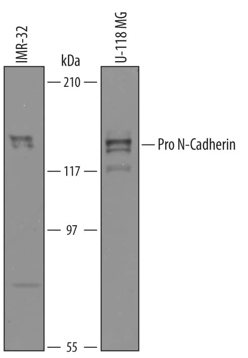
Detection of Human Pro-N-Cadherin by Western Blot.
Western blot shows lysates of IMR-32 human neuroblastoma cell line and U-118-MG human glioblastoma/astrocytoma cell line. PVDF membrane was probed with 1 µg/mL of Sheep Anti-Human N-Cadherin Propeptide Antigen Affinity-purified Polyclonal Antibody (Catalog # AF1388) followed by HRP-conjugated Anti-Sheep IgG Secondary Antibody (Catalog # HAF016). Pro-N-Cadherin was detected at approximately 130 kDa (as indicated). This experiment was conducted under reducing conditions and using Immunoblot Buffer Group 8.N‑Cadherin in Human Heart.
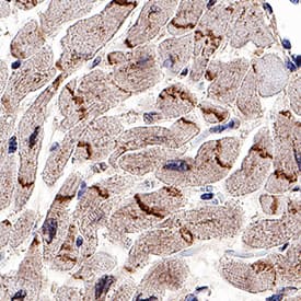
N-Cadherin was detected in immersion fixed paraffin-embedded sections of human heart using Sheep Anti-Human N-Cadherin Antigen Affinity-purified Polyclonal Antibody (Catalog # AF1388) at 1 µg/mL overnight at 4 °C. Tissue was stained using the Sheep IgG VisUCyte HRP Polymer Antibody (Catalog # VC006). Before incubation with the primary antibody, tissue was subjected to heat-induced epitope retrieval using Antigen Retrieval Reagent-Basic (Catalog # CTS013). Tissue was stained using DAB (brown) and counterstained with hematoxylin (blue). Specific staining was localized to intercalated disks between the myocardial cells. Staining was performed using our protocol for IHC Staining with VisUCyte HRP Polymer Detection Reagents.Applications for Human N-Cadherin Propeptide Antibody
Immunohistochemistry
Sample: Immersion fixed paraffin-embedded sections of human heart
Western Blot
Sample: IMR‑32 human neuroblastoma cell line and U‑118‑MG human glioblastoma/astrocytoma cell line
Formulation, Preparation, and Storage
Purification
Reconstitution
Formulation
Shipping
Stability & Storage
- 12 months from date of receipt, -20 to -70 °C as supplied.
- 1 month, 2 to 8 °C under sterile conditions after reconstitution.
- 6 months, -20 to -70 °C under sterile conditions after reconstitution.
Background: N-Cadherin
N‑Cadherin (Neural Cadherin; also CD325 and Cadherin-2) is a 130‑135 kDa member of the "classical" (or type I) cadherin subfamily, cadherin superfamily of proteins. It is expressed on multiple cell types, including neurons, fibroblasts, Schwann cells, endothelial cells and hepatic stellate cells. N‑Cadherin mediates homotypic binding, either in cis (same cell) or trans (adjacent cell). proN‑Cadherin is expressed as an 881 amino acid (aa) type I transmembrane glycoprotein. It may be initially inserted into the ER, where the 15‑20 kDa prodomain (aa 26‑159) is cleaved by proprotein convertase, and the mature molecule is transported to the surface. Alternatively, on neurons, proN‑Cadherin may first appear on the surface, with cleavage occurring at the time of synaptogenesis. Cleavage appears necessary for homophilic interaction as presence of the prodomain is suggested to negatively regulate oligomer formation. Over the entire prodomain, the human N‑Cadherin proregion shares 87% aa identity with the mouse N‑Cadherin proregion.
Long Name
Alternate Names
Gene Symbol
UniProt
Additional N-Cadherin Products
Product Documents for Human N-Cadherin Propeptide Antibody
Product Specific Notices for Human N-Cadherin Propeptide Antibody
For research use only
