Human Neuropilin-1 Antibody
R&D Systems, part of Bio-Techne | Catalog # AF3870

Key Product Details
Validated by
Species Reactivity
Validated:
Cited:
Applications
Validated:
Cited:
Label
Antibody Source
Product Specifications
Immunogen
Phe22-Lys644
Accession # NP_001019799
Specificity
Clonality
Host
Isotype
Endotoxin Level
Scientific Data Images for Human Neuropilin-1 Antibody
Detection of Human Neuropilin‑1 by Western Blot.
Western blot shows lysates of MDA-MB-231 human breast cancer cell line and human placenta tissue. PVDF membrane was probed with 1 µg/mL of Sheep Anti-Human Neuropilin-1 Antigen Affinity-purified Polyclonal Antibody (Catalog # AF3870) followed by HRP-conjugated Anti-Sheep IgG Secondary Antibody (Catalog # HAF016). A specific band was detected for Neuropilin-1 at approximately 130 kDa (as indicated). This experiment was conducted under reducing conditions and using Immunoblot Buffer Group 1.Detection of Neuropilin‑1 in HUVEC Human Cells by Flow Cytometry.
HUVEC human umbilical vein endothelial cells were stained with Sheep Anti-Human Neuropilin-1 Antigen Affinity-purified Polyclonal Antibody (Catalog # AF3870, filled histogram) or control antibody (Catalog # 5-001-A, open histogram), followed by NorthernLights™ 637-conjugated Anti-Sheep IgG Secondary Antibody (Catalog # NL011).Neuropilin‑1 in PC-3 and HLDM-2 Human Cell Lines.
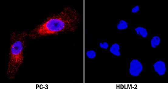
Neuropilin-1 was detected in immersion fixed PC-3 human prostate cancer cell line (positive staining) and HLDM-2 human Hodgkin's lymphoma cell line (negative staining) using Sheep Anti-Human Neuropilin-1 Antigen Affinity-purified Polyclonal Antibody (Catalog # AF3870) at 5 µg/mL for 3 hours at room temperature. Cells were stained using the NorthernLights™ 557-conjugated Anti-Sheep IgG Secondary Antibody (red; Catalog # NL010) and counterstained with DAPI (blue). Specific staining was localized to cytoplasm. View our protocol for Fluorescent ICC Staining of Cells on Coverslips.Applications for Human Neuropilin-1 Antibody
Blockade of Receptor-ligand Interaction
CyTOF-ready
Flow Cytometry
Sample: HUVEC human umbilical vein endothelial cells
Immunocytochemistry
Sample: Immersion fixed PC-3 human prostate cancer cell line and HLDM-2 human Hodgkin's lymphoma cell line
Immunohistochemistry
Sample: Immersion fixed paraffin-embedded sections of human ovarian and pancreatic cancer tissue
Western Blot
Sample: MDA‑MB‑231 human breast cancer cell line and human placenta tissue
Reviewed Applications
Read 5 reviews rated 3.8 using AF3870 in the following applications:
Formulation, Preparation, and Storage
Purification
Reconstitution
Formulation
Shipping
Stability & Storage
- 12 months from date of receipt, -20 to -70 °C as supplied.
- 1 month, 2 to 8 °C under sterile conditions after reconstitution.
- 6 months, -20 to -70 °C under sterile conditions after reconstitution.
Background: Neuropilin-1
Neuropilin-1 (Npn-1, previously neuropilin; also CD304/BDCA4) is a 130-140 kDa type I transmembrane (TM) glycoprotein that regulates axon guidance and angiogenesis (1-4). The full-length 923 amino acid (aa) human Npn-1 contains a 623 aa extracellular domain (ECD) that shows 92-95% aa identity with mouse, rat, bovine, and canine Npn-1 (3, 4). The ECD contains two N-terminal CUB domains (termed a1a2), two domains with homology to coagulation factors V and VIII (b1b2) and a MAM (meprin) domain (c). C-terminally divergent splice variants with 704, 644, 609, and 551 aa lack the MAM and TM domains and are demonstrated or presumed to be soluble antagonists (1, 5-7). A 906 aa form lacks a TM segment, but secretion has not been found (8). The sema domains of Class III secreted semaphorins such as Sema3A bind Npn-1 a1a2 (9). Heparin, the heparin-binding forms of VEGF (VEGF165, VEGF-B, and VEGF-E), PlGF (PlGF‑2), and the C-terminus of Sema3 bind the b1b2 region (9, 10). Npn-1 and Npn-2 share 48% aa identity within the ECD and can form homo- and hetero-oligomers via interaction of their MAM domains (1). Neuropilins show partially overlapping expression in neuronal and endothelial cells during development (1, 2). Both neuropilins act as co-receptors with plexins, mainly plexin A3 and A4, to bind class III semaphorins that mediate axon repulsion (11). However, only Npn-1 binds Sema3A, and only Npn-2 binds Sema3F (1). Both are co-receptors with VEGF R2 (also called KDR or Flk-1) for VEGF165 binding (1). Sema3A signaling can be blocked by VEGF165, which has higher affinity for Npn-1 (12). Npn-1 is preferentially expressed in arteries during development or those undergoing remodeling (1, 2). Npn-1 is also expressed on dendritic cells and mediates DC-induced T cell proliferation (13).
References
- Bielenberg, D.R. et al. (2006) Exp. Cell Res. 312:584.
- Gu, C. et al. (2003) Dev. Cell 5:45.
- He, Z. and M. Tessier-Lavigne (1997) Cell 90:739.
- Soker, S. et al. (1998) Cell 92:735.
- Gagnon, M.L. et al. (2000) Proc. Natl. Acad. Sci. USA 97:2573.
- Cackowski, F.C. et al. (2004) Genomics 84:82.
- Rossignol, M. et al. (2000) Genomics 70:211.
- Tao, Q. et al. (2003) Angiogenesis 6:39.
- Gu, C. et al. (2002) J. Biol. Chem. 277:18069.
- Mamluk, R. et al. (2002) J. Biol. Chem. 277:24818.
- Yaron, A. et al. (2005) Neuron 45:513.
- Narazaki, M. and G. Tosato (2006) Blood 107:3892.
- Tordjman, R. et al. (2002) Nat. Immunol. 3477.
Alternate Names
Gene Symbol
UniProt
Additional Neuropilin-1 Products
Product Documents for Human Neuropilin-1 Antibody
Product Specific Notices for Human Neuropilin-1 Antibody
This product or the use of this product is covered by U.S. Patents owned by The Regents of the University of California. This product is for research use only and is not to be used for commercial purposes. Use of this product to produce products for sale or for diagnostic, therapeutic or drug discovery purposes is prohibited. In order to obtain a license to use this product for such purposes, contact The Regents of the University of California.
U.S. Patent # 6,054,293, 6,623,738, and other U.S. and international patents pending.
For research use only


