Human NUB1 Antibody
R&D Systems, part of Bio-Techne | Catalog # AF7336

Key Product Details
Species Reactivity
Applications
Label
Antibody Source
Product Specifications
Immunogen
Arg513-Asn615
Accession # Q9Y5A7
Specificity
Clonality
Host
Isotype
Scientific Data Images for Human NUB1 Antibody
Detection of Human NUB1 by Western Blot.
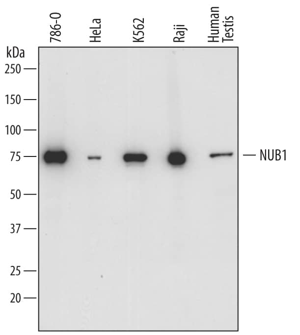
Western blot shows lysates of 786-O human renal cell adenocarcinoma cell line, HeLa human cervical epithelial carcinoma cell line, K562 human chronic myelogenous leukemia cell line, Raji human Burkitt's lymphoma cell line, and human testis tissue. PVDF membrane was probed with 0.5 µg/mL of Sheep Anti-Human NUB1 Antigen Affinity-purified Polyclonal Antibody (Catalog # AF7336) followed by HRP-conjugated Anti-Sheep IgG Secondary Antibody (HAF016). A specific band was detected for NUB1 at approximately 75 kDa (as indicated). This experiment was conducted under reducing conditions and using Immunoblot Buffer Group 1.Detection of Human NUB1 by Simple WesternTM.
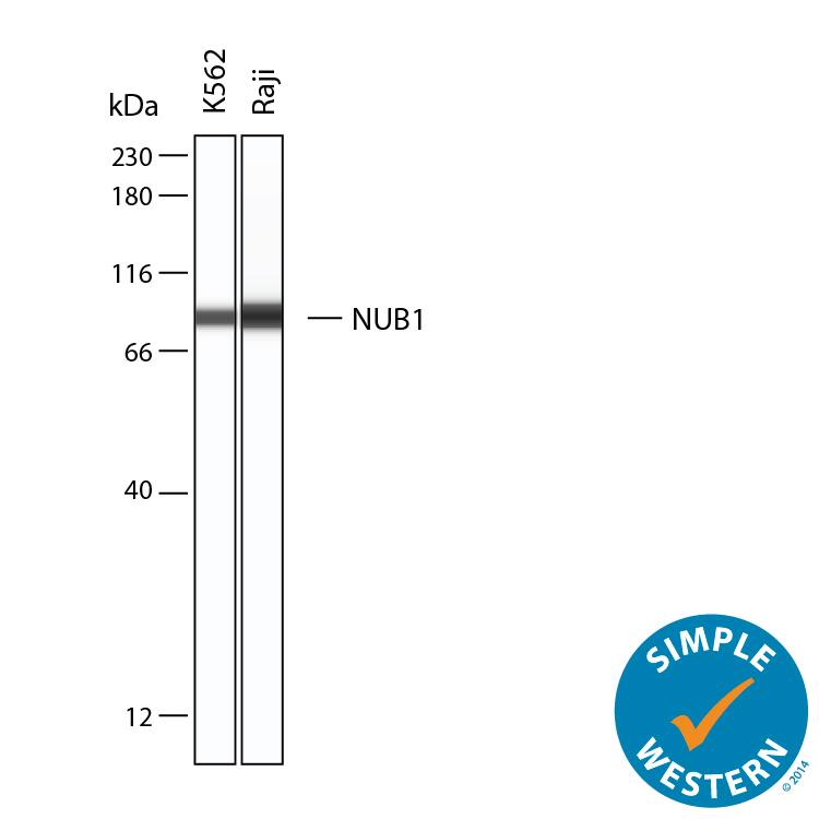
Simple Western lane view shows lysates of K562 human chronic myelogenous leukemia cells, and Raji human Burkitt's lymphoma cells, loaded at 0.2 mg/mL. A specific band was detected for NUB1 at approximately 89 kDa (as indicated) using 20 µg/mL of Sheep Anti-Human NUB1 Antigen Affinity-purified Polyclonal Antibody (Catalog # AF7336) followed by 1:50 dilution of HRP-conjugated Anti-Sheep IgG Secondary Antibody (Catalog # HAF016). This experiment was conducted under reducing conditions and using the 12-230 kDa separation system.Applications for Human NUB1 Antibody
Simple Western
Sample: K562 human chronic myelogenous leukemia cells, and Raji human Burkitt's lymphoma cells
Western Blot
Sample: 786‑O human renal cell adenocarcinoma cell line, HeLa human cervical epithelial carcinoma cell line, K562 human chronic myelogenous leukemia cell line, Raji human Burkitt's lymphoma cell line, and human testis tissue
Formulation, Preparation, and Storage
Purification
Reconstitution
Formulation
Shipping
Stability & Storage
- 12 months from date of receipt, -20 to -70 °C as supplied.
- 1 month, 2 to 8 °C under sterile conditions after reconstitution.
- 6 months, -20 to -70 °C under sterile conditions after reconstitution.
Background: NUB1
NUB1 (NEDD8 Ultimate Buster-1; also NY-REN18) is a 68-74 kDa, TNF-alpha - and IFN-inducible protein that is found intracellularly in a wide variety of cells. It is reported to be a down-regulator of the NEDD8 conjugation system. NEDD8 is a ubiquitin-like protein that links to cullin family of proteins. Cullin family proteins are associated with large SCF/E3 ligase complexes. In the case of cullin-1, the activities of its SCF-associated complex include the ubiquitination of I kappaB alpha, beta-catenin and p21. Ubiquitination does not occur, however, unless cullin-1 is conjugated to NEDD8, making NEDD8 a key regulator of ubiquitination. NUB1 downregulates ubiquitination by binding, and targeting, both free and conjugated NEDD8 to the proteosome. Human NUB1 is 615 amino acids (aa) in length. It contains two coiled-coil regions (aa 36-70 and 152-203) and three UBA domains (aa 374-413, 424-470 and 489-529). There are three potential isoform variants. One possesses an alternative start site 24 aa upstream of the standard site, a second shows a deletion of aa 452-465, and a third shows a combination of the first two variants. Over aa 513-615, human NUB1 shares 84% aa sequence identity with mouse NUB1.
Long Name
Alternate Names
Gene Symbol
UniProt
Additional NUB1 Products
Product Documents for Human NUB1 Antibody
Product Specific Notices for Human NUB1 Antibody
For research use only

