Human OX40/TNFRSF4 Antibody
R&D Systems, part of Bio-Techne | Catalog # AF3388


Key Product Details
Validated by
Species Reactivity
Validated:
Cited:
Applications
Validated:
Cited:
Label
Antibody Source
Product Specifications
Immunogen
Leu29-Ala216
Accession # P43489
Specificity
Clonality
Host
Isotype
Scientific Data Images for Human OX40/TNFRSF4 Antibody
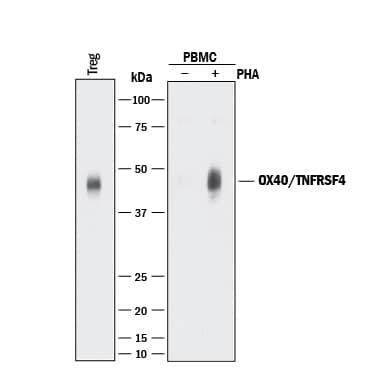
Detection of Human OX40/TNFRSF4 by Western Blot.
Western blot shows lysates of human T regulatory cells (Tregs) and human peripheral blood mononuclear cells (PBMCs) untreated (-) or treated (+) with 1 µg/mL PHA for 5 days. PVDF membrane was probed with 0.5 µg/mL of Sheep Anti-Human OX40/TNFRSF4 Antigen Affinity-purified Polyclonal Antibody (Catalog # AF3388) followed by HRP-conjugated Anti-Sheep IgG Secondary Antibody (Catalog # HAF016). A specific band was detected for OX40/TNFRSF4 at approximately 45 kDa (as indicated). This experiment was conducted under reducing conditions and using Immunoblot Buffer Group 1.Detection of OX40/TNFRSF4 in Human T Cells by Flow Cytometry.
Human T cells were treated for 3-5 days with 1 µg/mL PHA then stained with Sheep Anti-Human OX40/ TNFRSF4 Antigen Affinity-purified Polyclonal Antibody (Catalog # AF3388, filled histogram) or control antibody (Catalog # 5-001-A, open histogram), followed by NorthernLights™ 557-conjugated Anti-Sheep IgG Secondary Antibody (Catalog # NL010).OX40/TNFRSF4 in Human Tonsil.
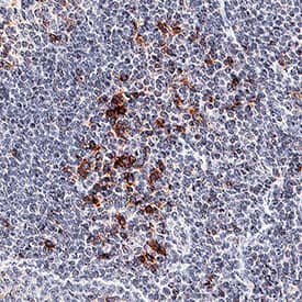
OX40/TNFRSF4 was detected in immersion fixed paraffin-embedded sections of human tonsil using Sheep Anti-Human OX40/TNFRSF4 Antigen Affinity-purified Polyclonal Antibody (Catalog # AF3388) at 3 µg/mL for 1 hour at room temperature followed by incubation with the Anti-Sheep IgG VisUCyte™ HRP Polymer Antibody (Catalog # VC006). Tissue was stained using DAB (brown) and counterstained with hematoxylin (blue). Specific staining was localized to plasma membrane. View our protocol for IHC Staining with VisUCyte HRP Polymer Detection Reagents.Applications for Human OX40/TNFRSF4 Antibody
CyTOF-ready
Flow Cytometry
Sample: Human T cells treated with PHA
Immunohistochemistry
Sample:
Immersion fixed paraffin-embedded sections of human tonsil
Western Blot
Sample: Human T regulatory cells (Tregs) and human peripheral blood mononuclear cells (PBMCs) treated with PHA
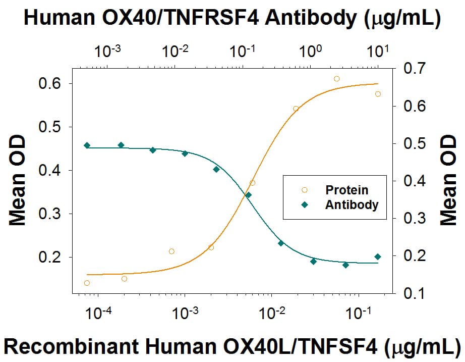
Neutralization
Reviewed Applications
Read 1 review rated 4 using AF3388 in the following applications:
Formulation, Preparation, and Storage
Purification
Reconstitution
Formulation
Shipping
Stability & Storage
- 12 months from date of receipt, -20 to -70 °C as supplied.
- 1 month, 2 to 8 °C under sterile conditions after reconstitution.
- 6 months, -20 to -70 °C under sterile conditions after reconstitution.
Background: OX40
OX40 (CD134; TNFRSF4) is a T cell costimulatory molecule of the TNF receptor superfamily that coordinates with other membrane-bound costimulators such as CD28, CD40, CD30, CD27 and 4-1BB (1‑3). OX40 is expressed on naïve CD4+ T cells only after engagement of the TCR by antigen presenting cells (APC; dendritic and B cells), and costimulation by CD40/CD40 ligand and CD28/B7. It is maximal at 2‑5 days post activation, or 4 hours post reactivation of memory T cells (3‑6). Human OX40 is a 48 kDa type I transmembrane glycoprotein with a 28 amino acid (aa) signal sequence, a 185 aa extracellular domain (ECD) that has four TNFR-Cys repeats and an O-glycosylated hinge region, a 20 aa transmembrane segment, and a 41 aa cytoplasmic domain (3). The ECD of human OX40 shows 71%, 68%, 67%, 64% and 64% aa identity with feline, canine, rabbit, mouse and rat OX40 ECD, respectively. Engagement of OX40 on activated CD4+ T cells by OX40 ligand on activated dendritic cells promotes T cell survival and proliferation, prolongs the immune response, and enhances the number of cells making the transition from effector to memory T cells (1‑6). OX40 signal transduction includes binding TNF receptor-associated factors (TRAFs), and activating NF kappaB and PI3 kinase to enhance expression of cytokines, antiapoptotic Bcl-2 family members, survivin and the chemokine receptor CXCR5 (5‑8). CXCR5 promotes T cell migration to germinal centers to deliver B cell help (5). Studies using knockout or transgenic mice, and agonistic or blocking antibodies, show that OX40/OX40L interaction is critical for establishing or reactivating memory T cells and breaking immune tolerance (9). Blockade of OX40 engagement is efficacious in animal models of allergic airway inflammation, graft-versus-host disease and autoimmune disease (10‑14).
References
- Salek-Ardakani, S. and M. Croft (2006) Vaccine 24:872.
- Hori, T. (2006) Int. J. Hematol. 83:17.
- Latza, U. et al. (1994) Eur. J. Immunol. 24:677.
- Murata, K. et al. (2000) J. Exp. Med. 191:365.
- Fillatreau, S and D. Gray (2003) J. Exp. Med. 197:195.
- Gramaglia, I. et al. (1998) J. Immunol. 161:6510.
- Rogers, P.R. et al. (2001) Immunity 15:445.
- Song, J. et al. (2005) Immunity 22:621.
- Bansal-Pakala, P. et al. (2001) Nat. Med. 7:907.
- Salek-Ardakani, S. et al. (2003) J. Exp. Med. 198:315.
- Jember, A. G. et al. (2001) J. Exp. Med. 193:387.
- Demirci, G. et al. (2004) J. Immunol. 172:1691.
- Blazar, B.R. et al. (2003) Blood 101:3741.
- Higgins, L.M. et al. (1999) J. Immunol. 162:486.
Alternate Names
Gene Symbol
UniProt
Additional OX40 Products
Product Documents for Human OX40/TNFRSF4 Antibody
Product Specific Notices for Human OX40/TNFRSF4 Antibody
For research use only


