Human P-Cadherin PE-conjugated Antibody
R&D Systems, part of Bio-Techne | Catalog # FAB861P


Key Product Details
Validated by
Species Reactivity
Applications
Label
Antibody Source
Product Specifications
Immunogen
Asp108-Gly654
Accession # CAA45177
Specificity
Clonality
Host
Isotype
Scientific Data Images for Human P-Cadherin PE-conjugated Antibody
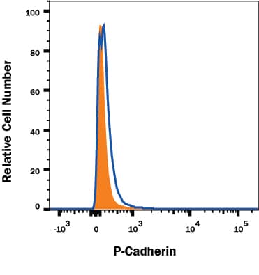
Detection of P‑Cadherin in A431 Human Cell Line by Flow Cytometry.
A431 human epithelial carcinoma cell line was stained with Mouse Anti-Human P-Cadherin PE-conjugated Monoclonal Antibody (Catalog # FAB861P, filled histogram) or isotype control antibody (Catalog # IC002P, open histogram). Cells were stained in a buffer containing Ca2+and Mg2+. View our protocol for Staining Membrane-associated Proteins.P-Cadherin Specificity is Shown by Flow Cytometry in Knockout Cell Line.
P-Cadherin knockout A431 human epithelial carcinoma cell line was stained with PE-conjugated Mouse Anti-Human P-Cadherin Monoclonal Antibody (Catalog # FAB861P, filled histogram) or isotype control antibody (Catalog # IC002P, open histogram). No staining in the P-Cadherin knockout A431 cell line was observed. Cells were stained in a buffer containing Ca2+ and Mg2+. View our protocol for Staining Membrane-associated Proteins.Applications for Human P-Cadherin PE-conjugated Antibody
Flow Cytometry
Sample: A431 human epithelial carcinoma cell line stained in a buffer containing Ca2+ and Mg2+
Knockout Validated
Formulation, Preparation, and Storage
Purification
Formulation
Shipping
Stability & Storage
- 12 months from date of receipt, 2 to 8 °C as supplied.
Background: P-Cadherin
Placental (P) - Cadherin (PCAD) is a member of the Cadherin family of cell adhesion molecules. Cadherins are calcium-dependent transmembrane proteins, which bind to one another in a homophilic manner. On their cytoplasmic side, they associate with the three catenins, alpha, beta, and gamma (plakoglobin). This association links the cadherin protein to the cytoskeleton. Without association with the catenins, the cadherins are non-adhesive. Cadherins play a role in development, specifically in tissue formation. They may also help to maintain tissue architecture in the adult. P-Cadherin is a classical cadherin molecule. Classical cadherins consist of a large extracellular domain which contains DXD and DXNDN repeats responsible for mediating calcium-dependent adhesion, a single-pass transmembrane domain, and a short carboxy-terminal cytoplasmic domain responsible for interacting with the catenins. Human P-Cadherin is an 829 amino acid (aa) protein with a 26 aa signal sequence and an 803 aa propeptide. The mature protein begins at aa 108 and has a 548 aa extracellular region, a 23 aa transmembrane region, and a 151 aa cytoplasmic region. The human and mouse mature PCAD proteins share 87% homology.
References
- Shimoyama, Y. et al. (1989) J. Cell Biol. 109:1787.
- Bussemakers, M.J.G. et al. (1993) Mol. Biol. Reports 17:123.
- Overduin, M. et al. (1995) Science 267:386.
- Takeichi, M. (1991) Science 251:1451.
- Nose, A. et al. (1987) EMBO J. 6:3655.
Long Name
Alternate Names
Gene Symbol
UniProt
Additional P-Cadherin Products
Product Documents for Human P-Cadherin PE-conjugated Antibody
Product Specific Notices for Human P-Cadherin PE-conjugated Antibody
For research use only
