Human PD-1 Biotinylated Antibody
R&D Systems, part of Bio-Techne | Catalog # BAF1086


Key Product Details
Validated by
Species Reactivity
Validated:
Cited:
Applications
Validated:
Cited:
Label
Antibody Source
Product Specifications
Immunogen
Leu25-Gln167
Accession # Q8IX89
Specificity
Clonality
Host
Isotype
Scientific Data Images for Human PD-1 Biotinylated Antibody
Detection of PD‑1 in Human PBMCs treated with PHA by Flow Cytometry.
Human peripheral blood mononuclear cells (PBMCs) either (A) untreated or (B) treated with 5 µg/mL PHA overnight were stained with Goat Anti-Human PD-1 Biotinylated Antigen Affinity-purified Polyclonal Antibody (Catalog # BAF1086) followed by Streptavidin-Phycoerythrin (Catalog # F0040) and Mouse Anti-Human CD3e APC-conjugated Monoclonal Antibody (Catalog # FAB100A). Quadrant markers were set based on control antibody staining (Catalog # BAF108). View our protocol for Staining Membrane-associated Proteins.Detection of Human PD-1 by Flow Cytometry
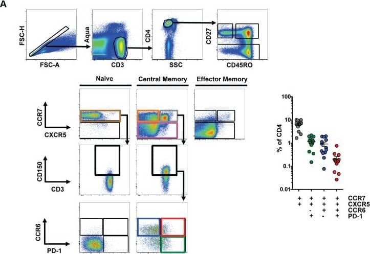
Characterization of peripheral TFH cells.(A) Left: Representative flow cytometry plots from HIV-uninfected PBMC showing the gating scheme for isolating T cell subsets for the T cell/B cell coculture assay. Isolated populations include naïve cells (brown), CM CCR7low (pink), CM CCR7highCXCR5low (orange), CM CCR7highCXCR5highCCR6lowPD-1high (green), CM CCR7highCXCR5highCCR6highPD-1low (blue) and CCR7highCXCR5highCCR6highPD-1high (red). Before gating on CCR6 and PD-1, cells were first gated on CD150high. Right: Scatter plot indicating the frequency of each population in HIV-uninfected subjects (n = 13). Cells were not gated on CD150 for phenotypic analysis. (B) Indicated CD4 T cell populations were cultured with autologous naïve B cells (CD19highCD27lowIgD−) in the presence of SEB for 12 days and Ig concentrations were measured from supernatants (n = 6). (C) Indicated CD4 T cell populations were cultured with autologous naïve B cells in the presence of SEB for 2 days and cytokine concentrations were measured from supernatants (n = 6). Horizontal lines indicate limit of detection. Significant differences were determined using the Friedman test with Dunn's multiple comparison post-test. *p<0.05; **p<0.01. Image collected and cropped by CiteAb from the following publication (https://pubmed.ncbi.nlm.nih.gov/24497824), licensed under a CC-BY license. Not internally tested by R&D Systems.Detection of Human PD-1 by Flow Cytometry
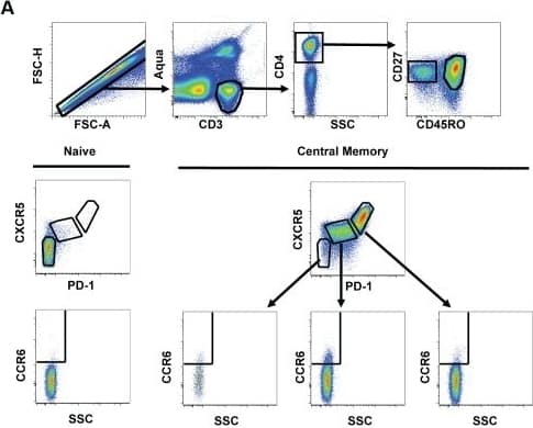
Relationship between pTFH cells and TFH cells in human tonsil.(A) Representative flow cytometry plots from HIV-uninfected, pediatric tonsils showing the gating scheme for determining the frequency of CCR6high cells in TFH (CXCR5highPD-1high) and non-TFH populations. (B) Bar graphs showing the frequency of CCR6high cells in TFH and non-TFH populations in human tonsils (n = 5). (C) Heatmap analysis of selected genes from RNA-seq data comparing pTFH cells (CXCR5highCCR6highPD-1high) from HIV-uninfected donors, pTFH cells from HIV-infected donors, non-TFH CD4 memory tonsil cells (CM CD57lowPD-1dimCCR7highCCR5lowCXCR4low), non-germinal center TFH tonsil cells (CM CD57lowPD-1highCCR7lowCXCR5high) and germinal center TFH tonsil cells (CM PD-1highCD57high) from HIV-uninfected donors. (D) Top: Comparison of MAF expression on CD4 T cells from blood or tonsil. Bottom: Geometric mean (MFI) of MAF expression in the indicated populations of central memory CD4 T cells normalized to MAF MFI in naïve CD4 T cells. Image collected and cropped by CiteAb from the following publication (https://pubmed.ncbi.nlm.nih.gov/24497824), licensed under a CC-BY license. Not internally tested by R&D Systems.Applications for Human PD-1 Biotinylated Antibody
Flow Cytometry
Sample: Human peripheral blood mononuclear cells (PBMCs) treated with PHA
Western Blot
Sample: Recombinant Human PD-1 Fc Chimera (Catalog # 1086-PD)
Human PD-1 Sandwich Immunoassay
Formulation, Preparation, and Storage
Purification
Reconstitution
Formulation
Shipping
Stability & Storage
- 12 months from date of receipt, -20 to -70 °C as supplied.
- 1 month, 2 to 8 °C under sterile conditions after reconstitution.
- 6 months, -20 to -70 °C under sterile conditions after reconstitution.
Background: PD-1
Programmed Death-1 (PD-1) is a type I transmembrane protein belonging to the CD28/CTLA-4 family of immunoreceptors that mediate signals for regulating immune responses (1). Members of the CD28/CTLA-4 family have been shown to either promote T cell activation (CD28 and ICOS) or downregulate T cell activation (CTLA-4 and PD-1) (2). PD-1 is expressed on activated T cells, B cells, myeloid cells, and on a subset of thymocytes. In vitro, ligation of PD-1 inhibits TCR-mediated T cell proliferation and production of IL-1, IL-4, IL-10, and IFN-gamma. In addition, PD-1 ligation also inhibits BCR mediated signaling. PD-1 deficient mice have a defect in peripheral tolerance and spontaneously develop autoimmune diseases (2, 3).
Two B7 family proteins, PD-L1 (also called B7-H1) and PD-L2 (also known as B7-DC), have been identified as PD-1 ligands. Unlike other B7 family proteins, both PD‑L1 and PD‑L2 are expressed in a wide variety of normal tissues including heart, placenta, and activated spleens (4). The wide expression of PD-L1 and PD-L2 and the inhibitor effects on PD-1 ligation indicate that PD-1 might be involved in the regulation of peripheral tolerance and may help prevent autoimmune diseases (2).
The human PD-1 gene encodes a 288 amino acid (aa) protein with a putative 20 aa signal peptide, a 148 aa extracellular region with one immunoglobulin-like V-type domain, a 24 aa transmembrane domain, and a 95 aa cytoplasmic region. The cytoplasmic tail contains two tyrosine residues that form the immuno-receptor tyrosine-based inhibitory motif (ITIM) and immunoreceptor tyrosine-based switch motif (ITSM) that are important in mediating PD-1 signaling. Mouse and human PD-1 share approximately 60% aa sequence identity (4).
References
- Ishida, Y. et al. (1992) EMBO J. 11:3887.
- Nishimura, H. and T. Honjo (2001) Trends in Immunol. 22:265.
- Latchman, Y. et al. (2001) Nature Immun. 2:261.
- Carreno, B.M. and M. Collins (2002) Annu. Rev. Immunol. 20:29.
Long Name
Alternate Names
Entrez Gene IDs
Gene Symbol
UniProt
Additional PD-1 Products
Product Documents for Human PD-1 Biotinylated Antibody
Product Specific Notices for Human PD-1 Biotinylated Antibody
For research use only

