Human PDCD4 Antibody
R&D Systems, part of Bio-Techne | Catalog # MAB7019

Key Product Details
Species Reactivity
Applications
Label
Antibody Source
Product Specifications
Immunogen
Lys212-His358
Accession # Q53EL6
Specificity
Clonality
Host
Isotype
Scientific Data Images for Human PDCD4 Antibody
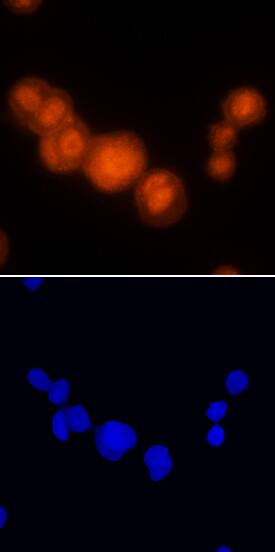
PDCD4 in MCF‑7 Human Cell Line.
PDCD4 was detected in immersion fixed MCF-7 human breast cancer cell line using Mouse Anti-Human PDCD4 Monoclonal Antibody (Catalog # MAB7019) at 25 µg/mL for 3 hours at room temperature. Cells were stained using the NorthernLights™ 557-conjugated Anti-Mouse IgG Secondary Antibody (red; Catalog # NL007) and counterstained with DAPI (blue). Specific staining was localized to nuclei and cytoplasm. View our protocol for Fluorescent ICC Staining of Cells on Coverslips.Applications for Human PDCD4 Antibody
Immunocytochemistry
Sample: Immersion fixed MCF-7 human breast cancer cell line
Formulation, Preparation, and Storage
Purification
Reconstitution
Formulation
Shipping
Stability & Storage
- 12 months from date of receipt, -20 to -70 °C as supplied.
- 1 month, 2 to 8 °C under sterile conditions after reconstitution.
- 6 months, -20 to -70 °C under sterile conditions after reconstitution.
Background: PDCD4
PDCD4 (Programmed cell death protein 4; also H731) is a 54-64 kDa member of the PDCD4 family of molecules. It is widely expressed, being found in mammary epithelium, CD34+ bone marrow progenitor cells, fibroblasts and keratinocytes. PDCD4 is both cytoplasmic and nuclear. In the cytoplasm, it blocks protein translation by binding to eIF4A, an act that dissociates eIF4G and mRNA from eIF4A. In the nucleus, it seems to block transcription of select genes, one of which is MAP4K1, a key enzyme in the AP-1-mediated transcription pathway. Human PDCD4 is 469 amino acids (aa) in length. It contains an NLS (aa 58-64), an MI domain (aa 163-284), another NLS (aa 241-250) and a second MI domain (aa 326-449). There are at least seven utilized Ser/Thr phosphorylation sites and one Tyr phosphorylation site. There are two potential splice forms, one that contains a deletion of aa 15-28, and a second that may show a three aa substitution for aa 2-15. Over aa 212-357, human PDCD4 shares 98% aa identity with mouse PDCD4.
Long Name
Alternate Names
Gene Symbol
UniProt
Additional PDCD4 Products
Product Documents for Human PDCD4 Antibody
Product Specific Notices for Human PDCD4 Antibody
For research use only
