Human R-Spondin 3 Antibody
R&D Systems, part of Bio-Techne | Catalog # MAB35001

Key Product Details
Species Reactivity
Applications
Label
Antibody Source
Product Specifications
Immunogen
Gln22-His272
Accession # Q9BXY4
Specificity
Clonality
Host
Isotype
Scientific Data Images for Human R-Spondin 3 Antibody
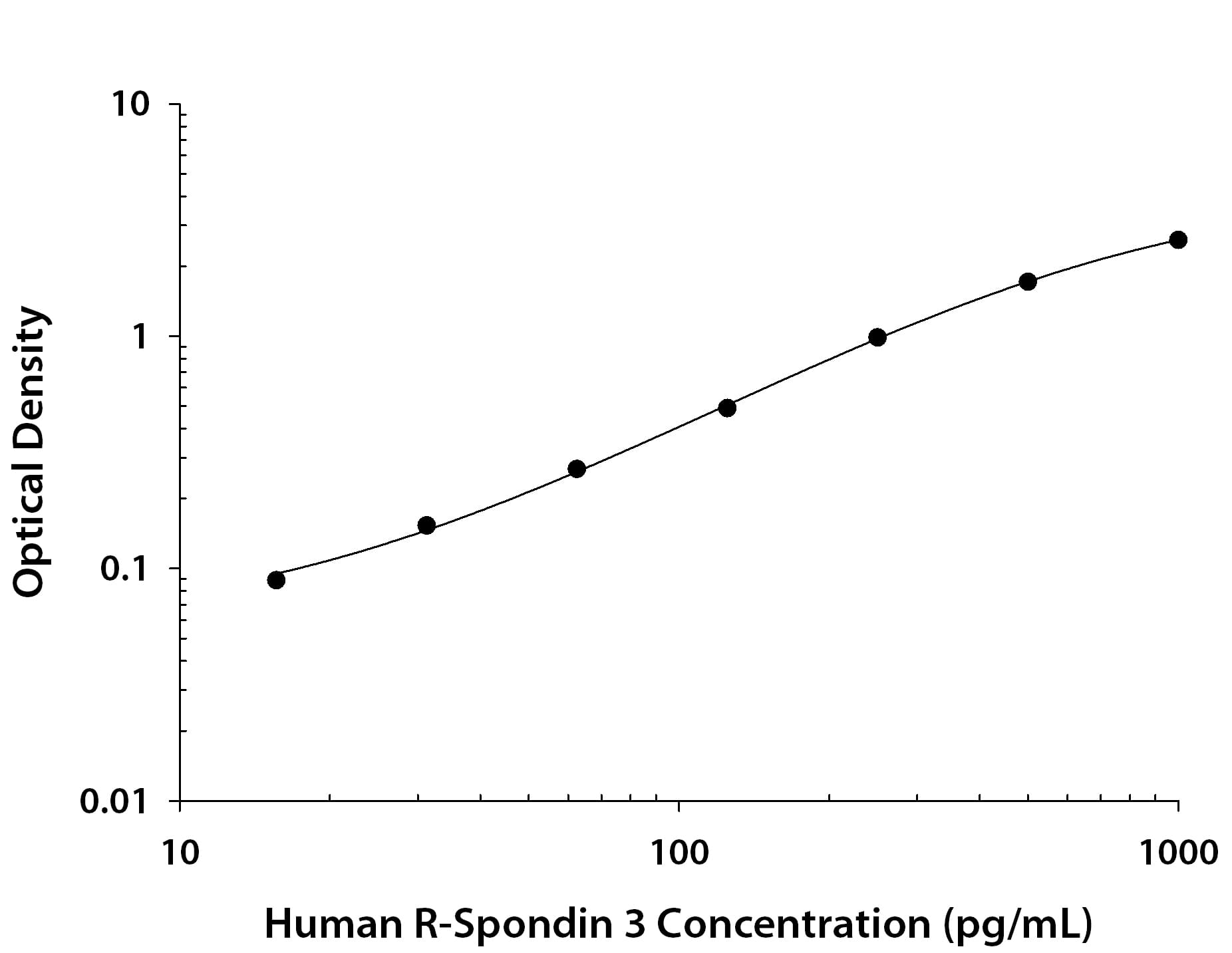
Human R-Spondin 3 ELISA Standard Curve.
Recombinant Human R-Spondin 3 protein was serially diluted 2-fold and captured by Mouse Anti-Human R-Spondin 3 Monoclonal Antibody (Catalog # MAB35001) coated on a Clear Polystyrene Microplate (DY990). Mouse Anti-Human R-Spondin 3 Monoclonal Antibody (MAB3500) was biotinylated and incubated with the protein captured on the plate. Detection of the standard curve was achieved by incubating Streptavidin-HRP (DY998) followed by Substrate Solution (DY999) and stopping the enzymatic reaction with Stop Solution (DY994).Applications for Human R-Spondin 3 Antibody
ELISA
This antibody functions as an ELISA capture antibody when paired with Mouse Anti-Human R‑Spondin 3 Monoclonal Antibody (Catalog # MAB3500).
This product is intended for assay development on various assay platforms requiring antibody pairs. We recommend the Human R-Spondin 3 DuoSet ELISA Kit (Catalog # DY3500) for convenient development of a sandwich ELISA.
Formulation, Preparation, and Storage
Purification
Reconstitution
Formulation
Shipping
Stability & Storage
- 12 months from date of receipt, -20 to -70 °C as supplied.
- 1 month, 2 to 8 °C under sterile conditions after reconstitution.
- 6 months, -20 to -70 °C under sterile conditions after reconstitution.
Background: R-Spondin 3
R-Spondin 3 (RSPO3, roof plate-specific spondin 3), also called cysteine-rich and single thrombospondin domain containing-1 (Cristin 1), is an ~31 kDa secreted protein that shares ~40% amino acid (aa) identity with the other three R-Spondin family members (1, 2). All are positive modulators of Wnt/ beta-catenin signaling, but each has a distinct expression pattern (1-4). Like other R-spondins,R-Spondin 3 contains two adjacent cysteine-rich furin-like domains (aa 35-135) with one potential N-glycosylation site (aa 36), followed by a thrombospondin (TSP-1) motif (aa 147-207) and a region rich in basic residues (aa 211-269). Only the furin-like domains are needed for beta-catenin stabilization (2). Within aa 21-209, human R-Spondin 3 shares 93%, 92%, 97%, 96% and 92% aa identity with mouse, rat, equine, bovine and canine R-Spondin 3, respectively. Potential isoforms of 279 and 297 aa diverge at aa 210 and 276, respectively (5). Mouse R-Spondin 3 is critical for development of the placental labyrinthine layer, probably by promoting VEGF expression and thus vascular development (6, 7). It is also essential for expression of the placenta-specific transcription factor, Gcm1. In the mouse embryo, R-Spondin 3 is often expressed by or located near endothelial cells (6). It is found in the roof plate, tail, somites, otic vesicles, cephalic mesoderm, truncus arteriosus, atrioventricular canal of the developing heart, and strongly but transiently in developing limbs (4, 7). R-Spondins regulate Wnt/ beta-catenin by competing with the Wnt antagonist DKK-1 for binding to the Wnt co-receptors LRP-6 and Kremen, reducing their DKK-1-mediated internalization (8, 9). Reports differ on whether R-Spondins bind LRP-6 directly (8-10). R-Spondin 3 has also been identified as an oncogene (11).
References
- Chen, J-Z. et al. (2002) Mol. Biol. Rep. 29:287.
- Kim, K.-A. et al. (2008) Mol. Biol. Cell 19:2588.
- Hendrickx, M. and L. Leyns (2008) Develop. Growth Differ. 50:229.
- Nam, J.-S. et al. (2007) Gene Expr. Patterns 7:306.
- Entrez Accession # EAW48114 and EAW48116.
- Kazanskaya, O. et al. (2008) Development 135:3655.
- Aoki, M. et al. (2007) Dev. Biol. 301:218.
- Binnerts, M.E. et al. (2007) Proc. Natl. Acad. Sci. USA 104:14700.
- Nam, J.-S. et al. (2006) J. Biol. Chem. 281:13247.
- Wei, Q. et al. (2007) J. Biol. Chem. 282:15903.
- Theodorou, V. et al. (2007) Nat. Genet. 6:759.
Long Name
Alternate Names
Gene Symbol
UniProt
Additional R-Spondin 3 Products
Product Documents for Human R-Spondin 3 Antibody
Product Specific Notices for Human R-Spondin 3 Antibody
For research use only
