Human RB1 Antibody
R&D Systems, part of Bio-Techne | Catalog # MAB6495

Key Product Details
Species Reactivity
Validated:
Cited:
Applications
Validated:
Cited:
Label
Antibody Source
Product Specifications
Immunogen
Lys240-Asn406
Accession # P06400
Specificity
Clonality
Host
Isotype
Scientific Data Images for Human RB1 Antibody
Detection of Human RB1 by Western Blot.
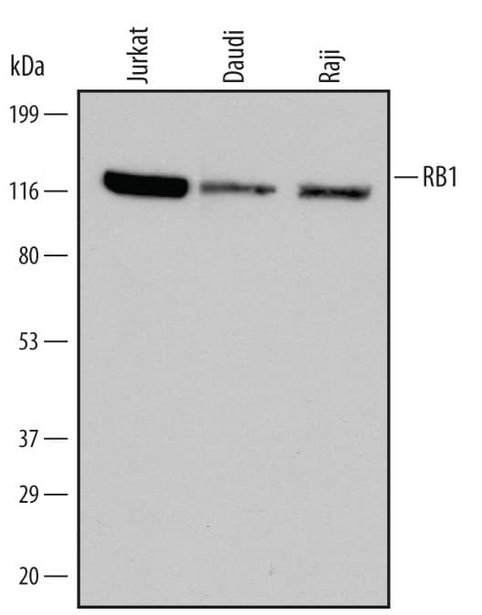
Western blot shows lysates of Jurkat human acute T cell leukemia cell line, Daudi human Burkitt's lymphoma cell line, and Raji human Burkitt's lymphoma cell line. PVDF Membrane was probed with 0.1 µg/mL of Human RB1 Monoclonal Antibody (Catalog # MAB6495) followed by HRP-conjugated Anti-Mouse IgG Secondary Antibody (Catalog # HAF007). A specific band was detected for RB1 at approximately 120 kDa (as indicated). This experiment was conducted under reducing conditions and using Immunoblot Buffer Group 1.RB1 in MCF‑7 Human Cell Line.
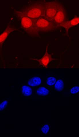
RB1 was detected in immersion fixed MCF-7 human breast cancer cell line using Human RB1 Monoclonal Antibody (Catalog # MAB6495) at 10 µg/mL for 3 hours at room temperature. Cells were stained using the NorthernLights™ 557-conjugated Anti-Mouse IgG Secondary Antibody (red, upper panel; Catalog # NL007) and counterstained with DAPI (blue, lower panel). Specific staining was localized to nuclei. View our protocol for Fluorescent ICC Staining of Cells on Coverslips.Detection of Human RB1 by Simple WesternTM.
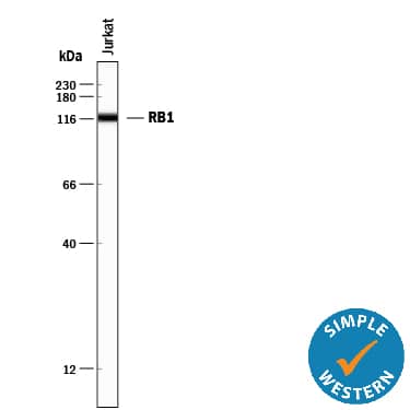
Simple Western lane view shows lysates of Jurkat human acute T cell leukemia cell line, loaded at 0.2 mg/mL. A specific band was detected for RB1 at approximately 120 kDa (as indicated) using 1 µg/mL of Mouse Anti-Human RB1 Monoclonal Antibody (Catalog # MAB6495). This experiment was conducted under reducing conditions and using the 12-230 kDa separation system.Applications for Human RB1 Antibody
Immunocytochemistry
Sample: Immersion fixed MCF-7 human breast cancer cell line
Simple Western
Sample: Jurkat human acute T cell leukemia cell line
Western Blot
Sample: Jurkat human acute T cell leukemia cell line, Daudi human Burkitt's lymphoma cell line, and Raji human Burkitt's lymphoma cell line
Formulation, Preparation, and Storage
Purification
Reconstitution
Formulation
Shipping
Stability & Storage
- 12 months from date of receipt, -20 to -70 °C as supplied.
- 1 month, 2 to 8 °C under sterile conditions after reconstitution.
- 6 months, -20 to -70 °C under sterile conditions after reconstitution.
Background: RB1
Retinoblastoma 1 protein (RB-1; also retinoblastoma-associated protein, pp110, and p105-Rb) is a 110 kDa tumor suppressor gene and member of the retinoblastoma protein family. Human RB-1 is 928 amino acids in length. The protein contains a Pocket domain (aa 373-771), which is comprised of two other domains, domain A (aa 373-573) and domain B (aa 640-771), and a “spacer” (aa 580-639). The Pocket domain binds to threonine-phosphorylated domain C (aa 771-928), which thereby prevents interaction with heterodimeric E2F/DP transcription factor complexes. Human RB-1 is 90% aa identical to mouse RB-1. RB-1 is expressed in the retina. The underphosphorylated, active form of RB-1 interacts with E2F1 and represses its transcription activity, leading to cell cycle arrest. Defects in RB-1 lead to the childhood cancer retinoblastoma.
Long Name
Alternate Names
Entrez Gene IDs
Gene Symbol
UniProt
Additional RB1 Products
Product Documents for Human RB1 Antibody
Product Specific Notices for Human RB1 Antibody
For research use only


