Human RBX1 Antibody
R&D Systems, part of Bio-Techne | Catalog # MAB11499

Key Product Details
Species Reactivity
Human
Applications
Immunocytochemistry, Immunohistochemistry, Simple Western, Western Blot
Label
Unconjugated
Antibody Source
Monoclonal Mouse IgG2B Clone # 1076128
Product Specifications
Immunogen
Spodoptera frugiperda, Sf 21 (baculovirus)-derived human CUL1/RBX1 Complex
Met1-His108
Accession # P62877
Met1-His108
Accession # P62877
Specificity
Detects human RBX1 in Western blots.
Clonality
Monoclonal
Host
Mouse
Isotype
IgG2B
Scientific Data Images for Human RBX1 Antibody
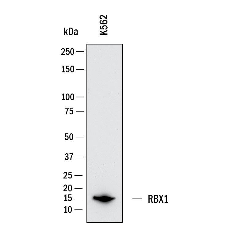
Detection of Human RBX1 by Western Blot.
Western Blot shows lysates of K562 human chronic myelogenous leukemia cell line. PVDF membrane was probed with 2 µg/ml of Mouse Anti-Human RBX1 Monoclonal Antibody (Catalog # MAB11499) followed by HRP-conjugated Anti-Mouse IgG Secondary Antibody (Catalog # HAF018). A specific band was detected for RBX1 at approximately 15 kDa (as indicated). This experiment was conducted under reducing conditions and using Western Blot Buffer Group 1.Detection of RBX1 in Prostate Tumor.
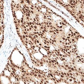
RBX1 was detected in immersion fixed paraffin-embedded sections of prostate tumor using Mouse Anti-Human RBX1 Monoclonal Antibody (Catalog # MAB11499) at 5 µg/ml for 1 hour at room temperature followed by incubation with the HRP-conjugated Anti-Mouse IgG Secondary Antibody (Catalog # HAF007) or the Anti-Mouse IgG VisUCyte™ HRP Polymer Antibody (Catalog # VC001). Before incubation with the primary antibody, tissue was subjected to heat-induced epitope retrieval using VisUCyte Antigen Retrieval Reagent-Basic (Catalog # VCTS021). Tissue was stained using DAB (brown) and counterstained with hematoxylin (blue). Specific staining was localized to the nucleus. View our protocol for Chromogenic IHC Staining of Paraffin-embedded Tissue Sections.Detection of RBX1 in MCF-7 cells (Positive) and Jurkat cells (Negative).
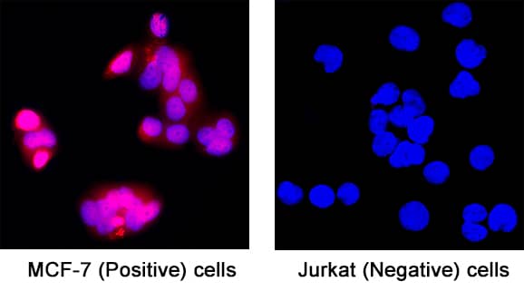
RBX1 was detected in fixed MCF-7 human breast cancer cell line (Positive) and absent in Jurkat human acute T cell leukemia cell line (Negative) using Mouse Anti-Human RBX1 Monoclonal Antibody (Catalog # MAB11499) at 8 µg/ml for 3 hours at room temperature. Cells were stained using the NorthernLights™ 557-conjugated Anti-Mouse IgG Secondary Antibody (red; Catalog # NL007) and counterstained with DAPI (blue). Specific staining was localized to the nucleus and cytoplasm. View our protocol for Fluorescent ICC Staining of Cells on Coverslips.Applications for Human RBX1 Antibody
Application
Recommended Usage
Immunocytochemistry
3-25 µg/mL
Sample: fixed MCF‑7 human breast cancer cell line (Positive) and absent in Jurkat human acute T cell leukemia cell line (Negative)
Sample: fixed MCF‑7 human breast cancer cell line (Positive) and absent in Jurkat human acute T cell leukemia cell line (Negative)
Immunohistochemistry
3-25 µg/mL
Sample: Immersion fixed paraffin-embedded sections of prostate tumor
Sample: Immersion fixed paraffin-embedded sections of prostate tumor
Simple Western
100 µg/mL
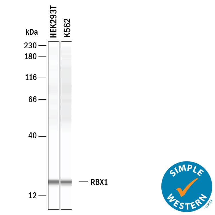
Sample: HEK293T human embryonic kidney cell line and K562 human chronic myelogenous leukemia cell line
Sample: HEK293T human embryonic kidney cell line and K562 human chronic myelogenous leukemia cell line
Western Blot
2 µg/mL
Sample: K562 human chronic myelongenous leukemia cell line
Sample: K562 human chronic myelongenous leukemia cell line
Formulation, Preparation, and Storage
Purification
Protein A or G purified from hybridoma culture supernatant
Reconstitution
Reconstitute at 0.5 mg/mL in sterile PBS. For liquid material, refer to CoA for concentration.
Formulation
Lyophilized from a 0.2 μm filtered solution in PBS with Trehalose. See Certificate of Analysis for details.
*Small pack size (-SP) is supplied either lyophilized or as a 0.2 µm filtered solution in PBS.
*Small pack size (-SP) is supplied either lyophilized or as a 0.2 µm filtered solution in PBS.
Shipping
Lyophilized product is shipped at ambient temperature. Liquid small pack size (-SP) is shipped with polar packs. Upon receipt, store immediately at the temperature recommended below.
Stability & Storage
Use a manual defrost freezer and avoid repeated freeze-thaw cycles.
- 12 months from date of receipt, -20 to -70 °C as supplied.
- 1 month, 2 to 8 °C under sterile conditions after reconstitution.
- 6 months, -20 to -70 °C under sterile conditions after reconstitution.
Background: RBX1
References
- Shao J, Feng Q, Jiang W, Yang Y, Liu Z, Li L, Yang W, Zou Y. E3 ubiquitin ligase RBX1 drives the metastasis of triple negative breast cancer through a FBXO45-TWIST1-dependent degradation mechanism. Aging (Albany NY). 2022 Jul 8;14(13):5493-5510. doi: 10.18632/aging.204163. Epub 2022 Jul 8. PMID: 35802537; PMCID: PMC9320552.
- Wei D, Sun Y. Small RING Finger Proteins RBX1 and RBX2 of SCF E3 Ubiquitin Ligases: The Role in Cancer and as Cancer Targets. Genes Cancer. 2010 Jul;1(7):700-7. doi: 10.1177/1947601910382776. PMID: 21103004; PMCID: PMC2983490.
Long Name
Ring-Box 1
Alternate Names
Ring Box 1, RNF75, ZYP Protein
Gene Symbol
RBX1
UniProt
Additional RBX1 Products
Product Documents for Human RBX1 Antibody
Product Specific Notices for Human RBX1 Antibody
For research use only
Loading...
Loading...
Loading...
Loading...


