Human STING/TMEM173 APC-conjugated Antibody
R&D Systems, part of Bio-Techne | Catalog # IC7169A


Conjugate
Catalog #
Key Product Details
Species Reactivity
Human
Applications
Intracellular Staining by Flow Cytometry
Label
Allophycocyanin (Excitation = 620-650 nm, Emission = 660-670 nm)
Antibody Source
Monoclonal Mouse IgG2B Clone # 723505
Product Specifications
Immunogen
E. coli-derived recombinant human STING/TMEM173
Ala215-Ser379
Accession # Q86WV6
Ala215-Ser379
Accession # Q86WV6
Specificity
Detects human STING/TMEM173 in direct ELISAs and Western blots.
Clonality
Monoclonal
Host
Mouse
Isotype
IgG2B
Scientific Data Images for Human STING/TMEM173 APC-conjugated Antibody
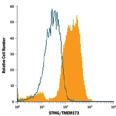
Detection of STING/TMEM173 in Human PBMC Monocytes by Flow Cytometry.
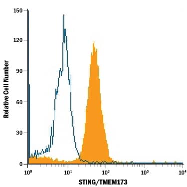
Human peripheral blood mononuclear cells (PBMC) monocytes were stained with Mouse Anti-Human STING/TMEM173 APC-conjugated Monoclonal Antibody (Catalog # IC7169A, filled histogram) or isotype control antibody (Catalog # IC0041A, open histogram). To facilitate intracellular staining, cells were fixed with Flow Cytometry Fixation Buffer (Catalog # FC004) and permeabilized with Flow Cytometry Permeabilization/Wash Buffer I (Catalog # FC005). View our protocol for Staining Intracellular Molecules.Detection of STING/TMEM173 in THP-1 Human Cell Line by Flow Cytometry.
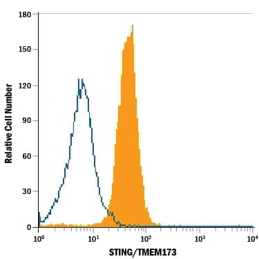
THP-1 human acute monocytic leukemia cell line was stained with Mouse Anti-Human STING/TMEM173 APC-conjugated Monoclonal Antibody (Catalog # IC7169A, filled histogram) or isotype control antibody (Catalog # IC0041A, open histogram). To facilitate intracellular staining, cells were fixed with Flow Cytometry Fixation Buffer (Catalog # FC004) and permeabilized with Flow Cytometry Permeabilization/Wash Buffer I (Catalog # FC005). View our protocol for Staining Intracellular Molecules.Detection of STING/TMEM173 in U937 Human Cell Line by Flow Cytometry.
U937 human histiocytic lymphoma cell line was stained with Mouse Anti-Human STING/TMEM173 APC-conjugated Monoclonal Antibody (Catalog # IC7169A, filled histogram) or isotype control antibody (Catalog # IC0041A, open histogram). To facilitate intracellular staining, cells were fixed with Flow Cytometry Fixation Buffer (Catalog # FC004) and permeabilized with Flow Cytometry Permeabilization/Wash Buffer I (Catalog # FC005). View our protocol for Staining Intracellular Molecules.Applications for Human STING/TMEM173 APC-conjugated Antibody
Application
Recommended Usage
Intracellular Staining by Flow Cytometry
10 µL/106 cells
Sample: Human peripheral blood mononuclear cells (PBMC) monocytes, THP‑1 human acute monocytic leukemia cell line, and U937 human histiocytic lymphoma cell line fixed with Flow Cytometry Fixation Buffer (Catalog # FC004) and permeabilized with Flow Cytometry Permeabilization/Wash Buffer I (Catalog # FC005)
Sample: Human peripheral blood mononuclear cells (PBMC) monocytes, THP‑1 human acute monocytic leukemia cell line, and U937 human histiocytic lymphoma cell line fixed with Flow Cytometry Fixation Buffer (Catalog # FC004) and permeabilized with Flow Cytometry Permeabilization/Wash Buffer I (Catalog # FC005)
Formulation, Preparation, and Storage
Purification
Protein A or G purified from hybridoma culture supernatant
Formulation
Supplied in a saline solution containing BSA and Sodium Azide.
Shipping
The product is shipped with polar packs. Upon receipt, store it immediately at the temperature recommended below.
Stability & Storage
Protect from light. Do not freeze.
- 12 months from date of receipt, 2 to 8 °C as supplied.
Background: STING/TMEM173
Long Name
Stimulator of Interferon Genes Protein/Transmembrane protein 173
Alternate Names
ERIS, MITA, MPYS, NET23, TMEM173
Gene Symbol
STING1
UniProt
Additional STING/TMEM173 Products
Product Documents for Human STING/TMEM173 APC-conjugated Antibody
Product Specific Notices for Human STING/TMEM173 APC-conjugated Antibody
For research use only
Loading...
Loading...
Loading...
Loading...
Loading...
Loading...

