Human TCF7/TCF1 Antibody
R&D Systems, part of Bio-Techne | Catalog # AF5596

Key Product Details
Species Reactivity
Human
Applications
Immunocytochemistry, Immunohistochemistry, Western Blot
Label
Unconjugated
Antibody Source
Polyclonal Goat IgG
Product Specifications
Immunogen
E. coli-derived recombinant human TCF7/TCF1
Met116-His233
Accession # P36402
Met116-His233
Accession # P36402
Specificity
Detects human TCF7/TCF1 in Western blots.
Clonality
Polyclonal
Host
Goat
Isotype
IgG
Scientific Data Images for Human TCF7/TCF1 Antibody
Detection of Human TCF7/TCF1 by Western Blot.
Western blot shows lysates of Jurkat human acute T cell leukemia cell line and MOLT-4 human acute lymphoblastic leukemia cell line. PVDF membrane was probed with 0.2 µg/mL of Goat Anti-Human TCF7/TCF1 Antigen Affinity-purified Polyclonal Antibody (Catalog # AF5596) followed by HRP-conjugated Anti-Goat IgG Secondary Antibody (Catalog # HAF019). Specific bands were detected for TCF7/TCF1 at approximately 60, 40, and 38 kDa (as indicated). This experiment was conducted under reducing conditions and using Immunoblot Buffer Group 1.TCF7/TCF1 in HCT‑116 Human Cell Line.
TCF7/TCF1 was detected in immersion fixed HCT-116 human colorectal carcinoma cell line using Human TCF7/TCF1 Antigen Affinity-purified Polyclonal Antibody (Catalog # AF5596) at 10 µg/mL for 3 hours at room temperature. Cells were stained using the NorthernLights™ 557-conjugated Anti-Goat IgG Secondary Antibody (red, upper panel; Catalog # NL001) and counterstained with DAPI (blue, lower panel). Specific staining was localized to nuclei. View our protocol for Fluorescent ICC Staining of Cells on Coverslips.TCF7/TCF1 in Human Thymus.
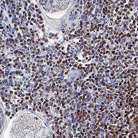
TCF7/TCF1 was detected in immersion fixed paraffin-embedded sections of human thymus using Goat Anti-Human TCF7/TCF1 Antigen Affinity-purified Polyclonal Antibody (Catalog # AF5596) at 3 µg/mL overnight at 4 °C. Tissue was stained using the Anti-Goat HRP-DAB Cell & Tissue Staining Kit (brown; Catalog # CTS008) and counterstained with hematoxylin (blue). Specific staining was localized to nuclei. View our protocol for Chromogenic IHC Staining of Paraffin-embedded Tissue Sections.Applications for Human TCF7/TCF1 Antibody
Application
Recommended Usage
Immunocytochemistry
5-15 µg/mL
Sample: Immersion fixed HCT-116 human colorectal carcinoma cell line
Sample: Immersion fixed HCT-116 human colorectal carcinoma cell line
Immunohistochemistry
3-15 µg/mL
Sample: Immersion fixed paraffin-embedded sections of human thymus
Sample: Immersion fixed paraffin-embedded sections of human thymus
Western Blot
0.2 µg/mL
Sample: Jurkat human acute T cell leukemia cell line and MOLT-4 human acute lymphoblastic leukemia cell line
Sample: Jurkat human acute T cell leukemia cell line and MOLT-4 human acute lymphoblastic leukemia cell line
Formulation, Preparation, and Storage
Purification
Antigen Affinity-purified
Reconstitution
Reconstitute at 0.2 mg/mL in sterile PBS. For liquid material, refer to CoA for concentration.
Formulation
Lyophilized from a 0.2 μm filtered solution in PBS with Trehalose. *Small pack size (SP) is supplied either lyophilized or as a 0.2 µm filtered solution in PBS.
Shipping
Lyophilized product is shipped at ambient temperature. Liquid small pack size (-SP) is shipped with polar packs. Upon receipt, store immediately at the temperature recommended below.
Stability & Storage
Use a manual defrost freezer and avoid repeated freeze-thaw cycles.
- 12 months from date of receipt, -20 to -70 °C as supplied.
- 1 month, 2 to 8 °C under sterile conditions after reconstitution.
- 6 months, -20 to -70 °C under sterile conditions after reconstitution.
Background: TCF7/TCF1
Long Name
Transcription Factor 7
Alternate Names
TCF1
Gene Symbol
TCF7
UniProt
Additional TCF7/TCF1 Products
Product Documents for Human TCF7/TCF1 Antibody
Product Specific Notices for Human TCF7/TCF1 Antibody
For research use only
Loading...
Loading...
Loading...
Loading...


