Human Thymosin beta4 Antibody
R&D Systems, part of Bio-Techne | Catalog # AF6796

Key Product Details
Species Reactivity
Validated:
Cited:
Applications
Validated:
Cited:
Label
Antibody Source
Product Specifications
Immunogen
Ser2-Ser44
Accession # P62328
Specificity
Clonality
Host
Isotype
Scientific Data Images for Human Thymosin beta4 Antibody
Detection of Human Thymosin beta4 by Western Blot.
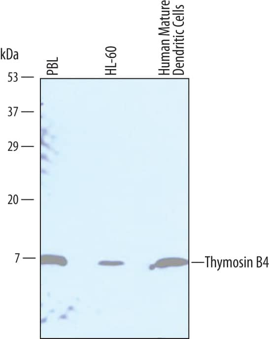
Western blot shows lysates of human peripheral blood lymphocytes (PBL), HL-60 human acute promyelocytic leukemia cell line, and human mature dendritic cells. PVDF Membrane was probed with 1 µg/mL of Sheep Anti-Human Thymosin beta4 Antigen Affinity-purified Polyclonal Antibody (Catalog # AF6796) followed by HRP-conjugated Anti-Sheep IgG Secondary Antibody (Catalog # HAF016). A specific band was detected for Thymosin beta4 at approximately 5 kDa (as indicated). This experiment was conducted under reducing conditions and using Immunoblot Buffer Group 1.Thymosin beta4 in HeLa Human Cell Line.
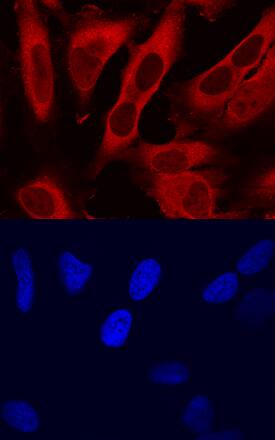
Thymosin beta4 was detected in immersion fixed HeLa human cervical epithelial carcinoma cell line using Sheep Anti-Human Thymosin beta4 Antigen Affinity-purified Polyclonal Antibody (Catalog # AF6796) at 10 µg/mL for 3 hours at room temperature. Cells were stained using the NorthernLights™ 557-conjugated Anti-Sheep IgG Secondary Antibody (red, upper panel; Catalog # NL010) and counterstained with DAPI (blue, lower panel). Specific staining was localized to cytoplasm. View our protocol for Fluorescent ICC Staining of Cells on Coverslips.Detection of Human Thymosin beta4 by Simple WesternTM.
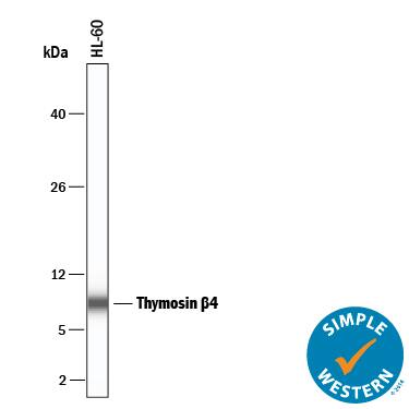
Simple Western lane view shows lysates of HL-60 human acute promyelocytic leukemia cell line, loaded at 0.2 mg/mL. A specific band was detected for Thymosin beta4 at approximately 8 kDa (as indicated) using 10 µg/mL of Sheep Anti-Human Thymosin beta4 Antigen Affinity-purified Polyclonal Antibody (Catalog # AF6796) followed by 1:50 dilution of HRP-conjugated Anti-Sheep IgG Secondary Antibody (HAF016). This experiment was conducted under reducing conditions and using the 2-40 kDa separation system.Applications for Human Thymosin beta4 Antibody
Immunocytochemistry
Sample: Immersion fixed HeLa human cervical epithelial carcinoma cell line
Simple Western
Sample: HL‑60 human acute promyelocytic leukemia cell line
Western Blot
Sample: Human peripheral blood lymphocytes (PBL), HL‑60 human acute promyelocytic leukemia cell line, and human mature dendritic cells
Formulation, Preparation, and Storage
Purification
Reconstitution
Formulation
Shipping
Stability & Storage
- 12 months from date of receipt, -20 to -70 °C as supplied.
- 1 month, 2 to 8 °C under sterile conditions after reconstitution.
- 6 months, -20 to -70 °C under sterile conditions after reconstitution.
Background: Thymosin beta 4
Thymosin beta 4 (T beta4; also TB4X and Fx) is a 5.0 kDa member of the beta-thymosin family of molecules. Members of this family range from 41-44 amino acids (aa) in length, and possess an isoelectric point that lies between pH 4.0-7.0 ( alpha-thymosins have values less than 4.0). Multiple cell types produce T beta4, either constitutively, or after stimulation. They include platelets, endothelial cells, neutrophils, astrocytes and macrophages. T beta4 is both a secreted and intracellular molecule. The secreted form contributes to wound healing and angiogenesis, and may act on ATPase. Intracellularly, it forms a 1:1 complex with G-actin and blocks F-actin polymerization. This regulates the availablility of actin monomers for filament formation and subsequent cell migration. Mature human T beta4 is 43 aa in length (aa 2-44). It contains an actin-binding site (aa 17-23), one acetylated Ser and five acetylated lysines (4; 12; 26; 32; 39) and one phosphorylation site at Thr23. T beta4 undergoes proteolytic processing to generate an N-terminal acetylated peptide (aa 2-5: SerAspLysPro). Mature human T beta4 is identical to mouse T beta14 in aa sequence, and it shares 74% aa identity with its human family member T beta10.
Additional Thymosin beta 4 Products
Product Documents for Human Thymosin beta4 Antibody
Product Specific Notices for Human Thymosin beta4 Antibody
For research use only


