Human TROP-2 Antibody
R&D Systems, part of Bio-Techne | Catalog # AF650

Key Product Details
Validated by
Species Reactivity
Validated:
Cited:
Applications
Validated:
Cited:
Label
Antibody Source
Product Specifications
Immunogen
Thr88-Thr274
Accession # P09758
Specificity
Clonality
Host
Isotype
Scientific Data Images for Human TROP-2 Antibody
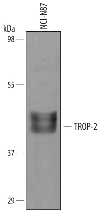
Detection of Human TROP-2 by Western Blot.
Western blot shows lysates of NCI-N87 human gastric carcinoma cell line. PVDF membrane was probed with 1 µg/mL of Goat Anti-Human TROP-2 Antigen Affinity-purified Polyclonal Antibody (Catalog # AF650) followed by HRP-conjugated Anti-Goat IgG Secondary Antibody (Catalog # HAF019). A specific band was detected for TROP-2 at approximately 45-50 kDa (as indicated). This experiment was conducted under reducing conditions and using Immunoblot Buffer Group 8.Detection of TROP‑2 in PC‑3 Human Cell Line by Flow Cytometry.
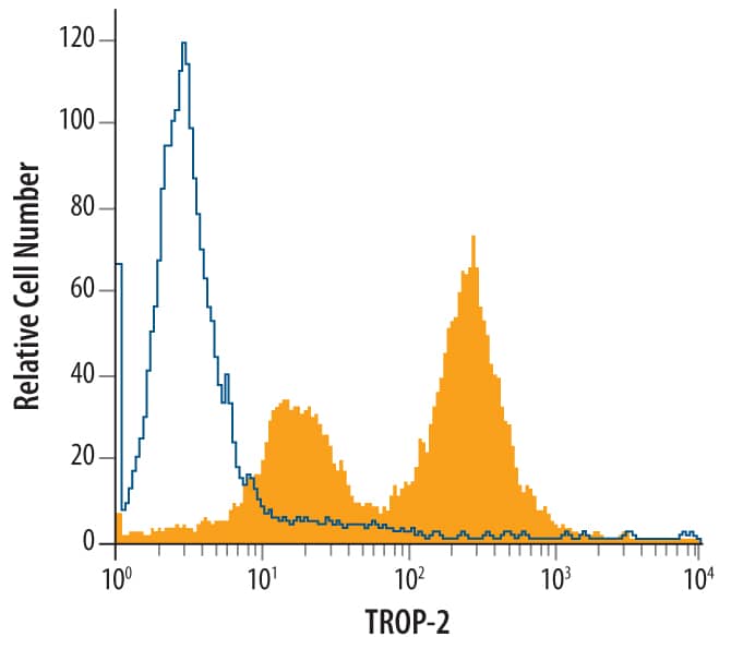
PC-3 human prostate cancer cell line was stained with Goat Anti-Human TROP-2 Antigen Affinity-purified Polyclonal Antibody (Catalog # AF650, filled histogram) or control antibody (Catalog # AB-108-C, open histogram), followed by Phycoerythrin-conjugated Anti-Goat IgG Secondary Antibody (Catalog # F0107).TROP-2 in Human Brain.
TROP-2 was detected in immersion fixed paraffin-embedded sections of human brain (frontal cortex) using Goat Anti-Human TROP-2 Antigen Affinity-purified Polyclonal Antibody (Catalog # AF650) at 10 µg/mL overnight at 4 °C. Before incubation with the primary antibody tissue was subjected to heat-induced epitope retrieval using Antigen Retrieval Reagent-Basic (Catalog # CTS013). Tissue was stained using the Anti-Goat HRP-DAB Cell & Tissue Staining Kit (brown; Catalog # CTS008) and counterstained with hematoxylin (blue). View our protocol for Chromogenic IHC Staining of Paraffin-embedded Tissue Sections.Applications for Human TROP-2 Antibody
CyTOF-ready
Flow Cytometry
Sample: PC-3 human prostate cancer cell line
Immunohistochemistry
Sample: Immersion fixed paraffin-embedded sections of human brain (cortex) subjected to Antigen Retrieval Reagent-Basic (Catalog # CTS013)
Western Blot
Sample: NCI‑N87 human gastric carcinoma cell line
Reviewed Applications
Read 1 review rated 4 using AF650 in the following applications:
Formulation, Preparation, and Storage
Purification
Reconstitution
Formulation
Shipping
Stability & Storage
- 12 months from date of receipt, -20 to -70 °C as supplied.
- 1 month, 2 to 8 °C under sterile conditions after reconstitution.
- 6 months, -20 to -70 °C under sterile conditions after reconstitution.
Background: TROP-2
Human TROP-2, also called tumor associated calcium signal transducer 2 (TACSTD2), GA733-1, gp50 and T16, is a type I cell surface glycoprotein that is highly expressed on human carcinomas. It was originally identified as an antigen present on human gastrointestinal tumors and is the second of two members of this family. The other family member is GA733-2, also called EpCAM, TROP-1, 17-1A, gp40 and KSA. The TROP-2 gene is unique in that it contains no introns. A study of these two genes suggested that TROP-2 was the result of a retroposition of the EpCAM gene. TROP-2 and EpCAM share approximately 49% amino acid identity and approximately 67% similarity. Human and mouse TROP-2 share 87% similarity. The human TROP-2 protein consists of a putative 26 amino acid (aa) signal sequence, a 248 aa extracellular domain, a 23 aa transmembrane region and a 26 aa cytoplasmic domain. TROP-2 is capable of transducing an intracellular calcium signal and may play a role in tumor growth. It also has adhesive functions.
References
- Linnenbach, A.J. et al. (1989) Proc. Natl. Acad. Sci. USA 86:27.
- Linnenbach, A.J. et al. (1993) Mol. Cell. Biol. 13:1507.
- Fornaro, M. et al. (1995) Int. J. Cancer 62:610.
- Ripani, E. et al. (1998) Int. J. Cancer 76:671.
- El Sewedy, T. et al. (1998) Int. J. Cancer 75:324.
Long Name
Alternate Names
Gene Symbol
UniProt
Additional TROP-2 Products
Product Documents for Human TROP-2 Antibody
Product Specific Notices for Human TROP-2 Antibody
For research use only
