Human VCAM-1/CD106 Antibody
R&D Systems, part of Bio-Techne | Catalog # BBA5

Key Product Details
Validated by
Biological Validation
Species Reactivity
Validated:
Human
Cited:
Human, Rat
Applications
Validated:
Adhesion Blockade, Immunocytochemistry, Immunoprecipitation, Western Blot
Cited:
Blocking, ELISA Development, ELISA Development (Capture), Flow Cytometry, Functional Assay, Immunocytochemistry, Immunohistochemistry, Immunoprecipitation, Neutralization, Western Blot
Label
Unconjugated
Antibody Source
Monoclonal Mouse IgG1 Clone # BBIG-V1
Product Specifications
Immunogen
Activated HUVEC human umbilical vein endothelial cells
Specificity
The antibody was screened using COS cells transfected with cDNAs for E-Selectin, VCAM-1, ICAM-1 and PECAM-1 and was shown to be specific for human VCAM-1/CD106.
Clonality
Monoclonal
Host
Mouse
Isotype
IgG1
Endotoxin Level
<0.10 EU per 1 μg of the antibody by the LAL method.
Scientific Data Images for Human VCAM-1/CD106 Antibody
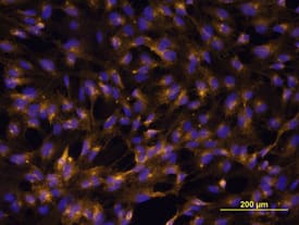
VCAM‑1/CD106 in HUVEC Cells.
VCAM-1/CD106 was detected in immersion fixed HUVEC human umbilical vein endothelial cells using Human VCAM-1/CD106 Monoclonal Antibody (Catalog # BBA5) at 10 µg/mL for 3 hours at room temperature. Cells were stained using the Northern-Lights™ 557-conjugated Anti-Mouse IgG Secondary Antibody (yellow; Catalog # NL007) and counter-stained with DAPI (blue). View our protocol for Fluorescent ICC Staining of Cells on Coverslips.This antibody is not suitable for labeling formalin-fixed, paraffin-embedded tissue./P>VCAM-1/CD106 in HUVEC cells treated with TNF alpha (positive) and untreated HUVEC cells (negative).
VCAM-1/CD106 was detected in immersion fixed HUVEC cells treated with TNF alpha (positive) and untreated HUVEC cells (negative) using Mouse Anti-Human VCAM-1/CD106 Monoclonal Antibody (Catalog # BBA5) at 8 µg/mL for 3 hours at room temperature. Cells were stained using the NorthernLights™ 557-conjugated Anti-Mouse IgG Secondary Antibody (red; Catalog # NL007) and counterstained with DAPI (blue). Specific staining was localized to plasma membrane. View our protocol for Fluorescent ICC Staining of Non-adherent Cells.Applications for Human VCAM-1/CD106 Antibody
Application
Recommended Usage
Adhesion Blockade
The adhesion of U937 human histiocytic lymphoma cells (5 x 10⁴ cells/well) to immobilized Recombinant Human VCAM-1/CD106 (Catalog # ADP5, 2.5 µg/mL, 100 µL/well) was maximally inhibited (80-100%) by 30 µg/mL of the antibody.
Immunocytochemistry
8-25 µg/mL
Sample: Immersion fixed HUVEC human umbilical vein endothelial cells, and immersion fixed HUVEC human umbilical vein endothelial cells activated with recombinant human TNF-alpha (Catalog # 210-TA). This antibody is not suitable for labeling formalin-fixed, paraffin-embedded tissue.
Sample: Immersion fixed HUVEC human umbilical vein endothelial cells, and immersion fixed HUVEC human umbilical vein endothelial cells activated with recombinant human TNF-alpha (Catalog # 210-TA). This antibody is not suitable for labeling formalin-fixed, paraffin-embedded tissue.
Immunoprecipitation
10 µg/mL
Sample: Radiolabelled activated HUVEC human umbilical vein endothelial cells, see our available Western blot detection antibodies
Sample: Radiolabelled activated HUVEC human umbilical vein endothelial cells, see our available Western blot detection antibodies
Western Blot
1 µg/mL
Sample: Recombinant Human VCAM‑1/CD106 (Catalog # 809-VR) under non-reducing conditions only
Sample: Recombinant Human VCAM‑1/CD106 (Catalog # 809-VR) under non-reducing conditions only
Reviewed Applications
Read 1 review rated 4 using BBA5 in the following applications:
Formulation, Preparation, and Storage
Purification
Protein A or G purified from hybridoma culture supernatant
Reconstitution
Sterile PBS to a final concentration of 0.5 mg/mL.
Formulation
Lyophilized from a 0.2 μm filtered solution in PBS with Trehalose.
Shipping
The product is shipped at ambient temperature. Upon receipt, store it immediately at the temperature recommended below.
Stability & Storage
Use a manual defrost freezer and avoid repeated freeze-thaw cycles.
- 12 months from date of receipt, -20 to -70 °C as supplied.
- 1 month, 2 to 8 °C under sterile conditions after reconstitution.
- 6 months, -20 to -70 °C under sterile conditions after reconstitution.
Background: VCAM-1/CD106
Long Name
Vascular Cell Adhesion Molecule 1
Alternate Names
CD106, VCAM1
Gene Symbol
VCAM1
Additional VCAM-1/CD106 Products
Product Documents for Human VCAM-1/CD106 Antibody
Product Specific Notices for Human VCAM-1/CD106 Antibody
For research use only
Loading...
Loading...
Loading...
Loading...


